Synaptophysin Immunoreactivity in the Brain of the P19 Tammar Wallaby (Notamacropus eugenii)
Introduction
The tammar wallaby (Notamacropus eugenii) is a small macropod originally found in Western and South Australia and on ten or more offshore islands. Please see the introduction for the P5 H&E atlas for details of tammar wallaby biology.
Synaptophysin (major synaptic vesicle protein p38) is a membrane glycoprotein that is found in presynaptic membranes of axons throughout the nervous system and in neuroendocrine cells. It may be important in the regulation of activity-dependent synapse formation (Tarsa and Goda, 2002), but its precise function is still uncertain. It is used here as a marker for development of axonal connections in the immature tammar nervous system.
Methods
The wallaby depicted in the series of colour plates was obtained from a breeding colony at the Australian National University (ANU) in the Australian Capital Territory (ACT).
All experimental procedures were approved by the Animal Ethics Experimentation Committee of the ANU, conform to NIH principles of laboratory animal care and were carried out according to the ethical guidelines of the National Health and Medical Research Council (Australia). The age of the pouch young animal was determined either directly by noting the elapsed time from the date of birth, which was designated P0, or from measurements of head-length and reference to a chart of head-lengths of animals of known age. This is accurate to within ±2 days.
The pouch-young tammar was anaesthetized by hypothermia and perfused with normal saline followed by Bouin’s fixative. The head was stored in 70% ethanol prior to embedding in paraffin and sectioning coronally at a thickness of 10 µm. Embedding, sectioning and immunohistochemistry all took place in the laboratory of Professor Jürgen Mai at the Heinrich Heine University in Düsseldorf. The sections were mounted on subbed glass slides. After immunostaining, the slides were coverslipped with DePeX.
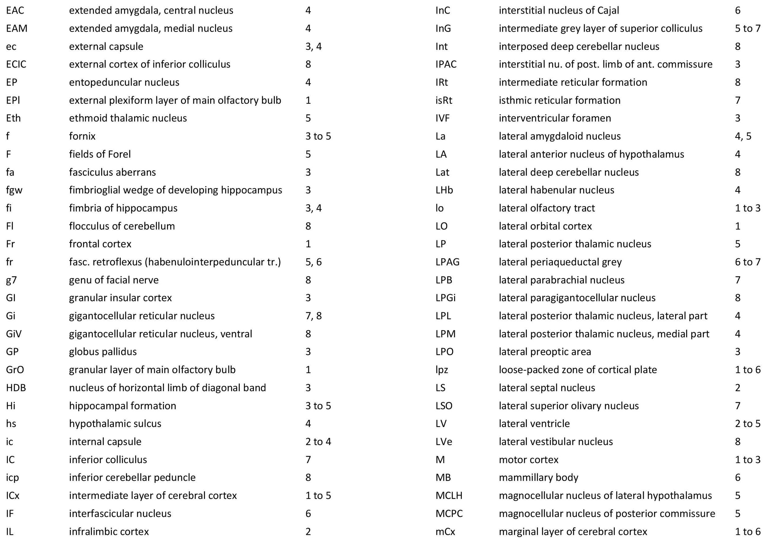
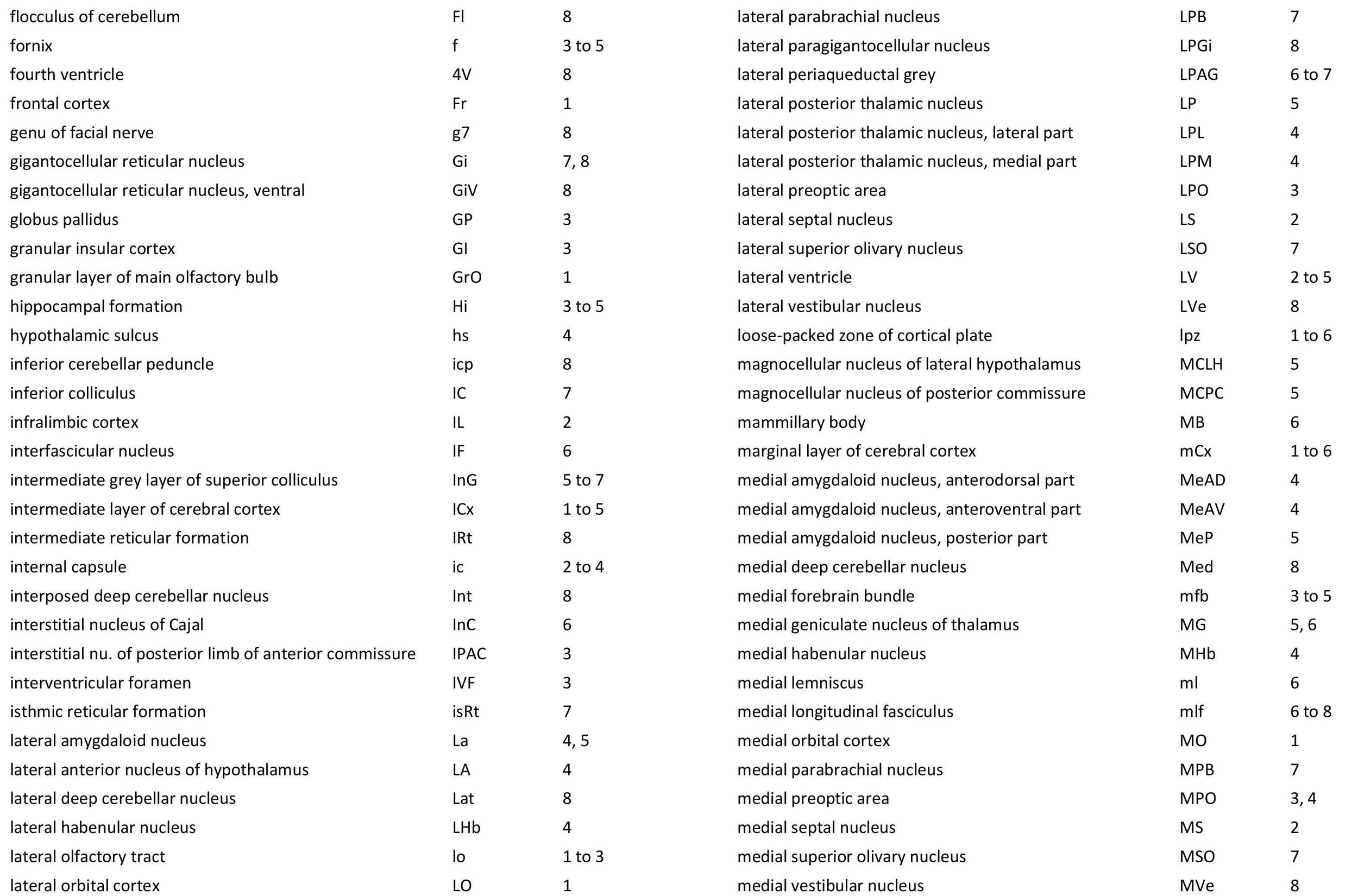
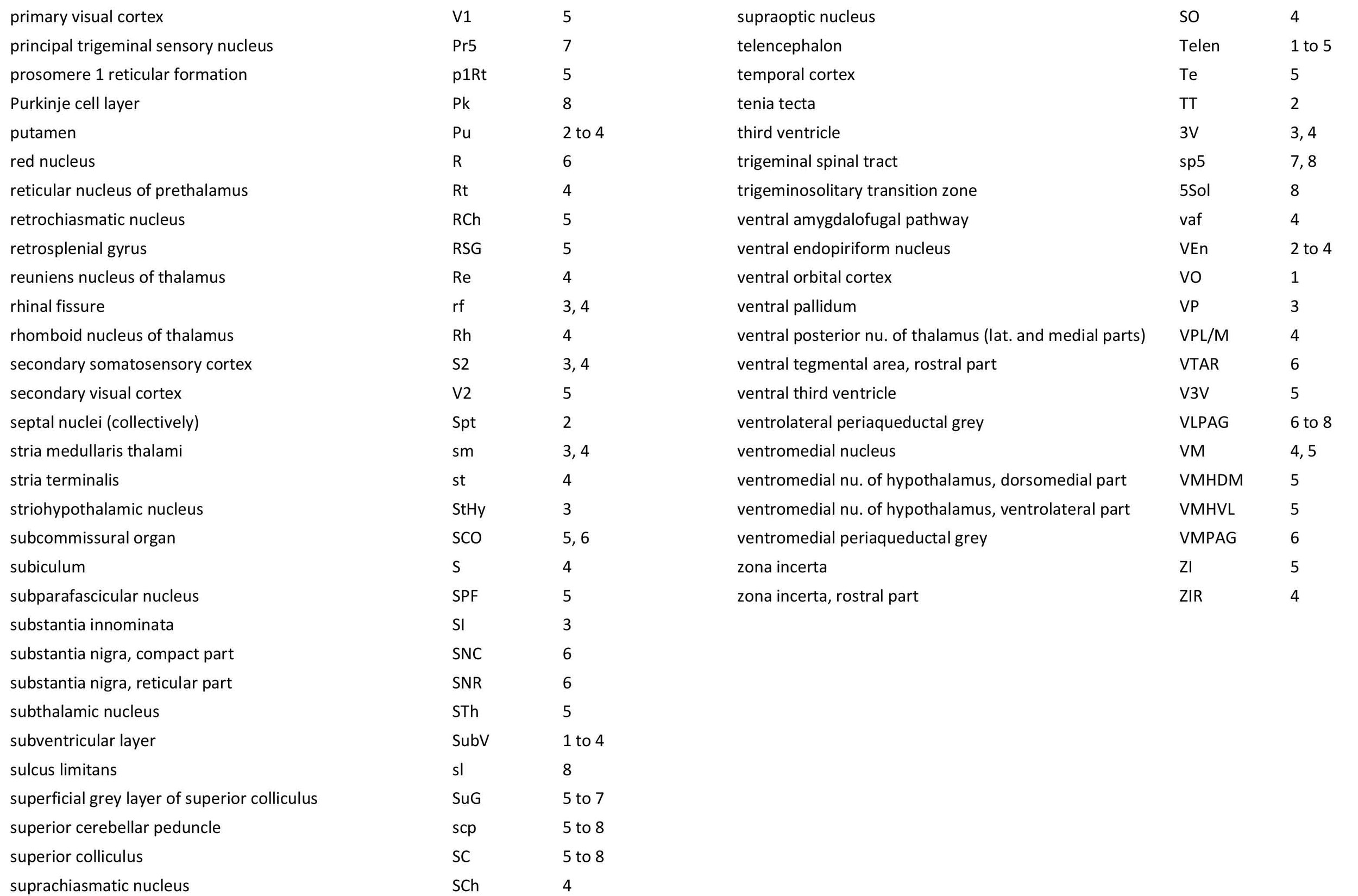
Each section was photographed with the aid of a Zeiss Axiophot photomicrographic system and a Canon EOS 400Dbody. Images were placed in Adobe Illustrator 2021 and delineated. Developmental regions (i.e. neuroepithelium) destined to give rise to adult structures have been denoted by the adult structure’s name with an asterisk (e.g. Cb* denotes the cerebellar developmental field of the rhombic lip).
Observations
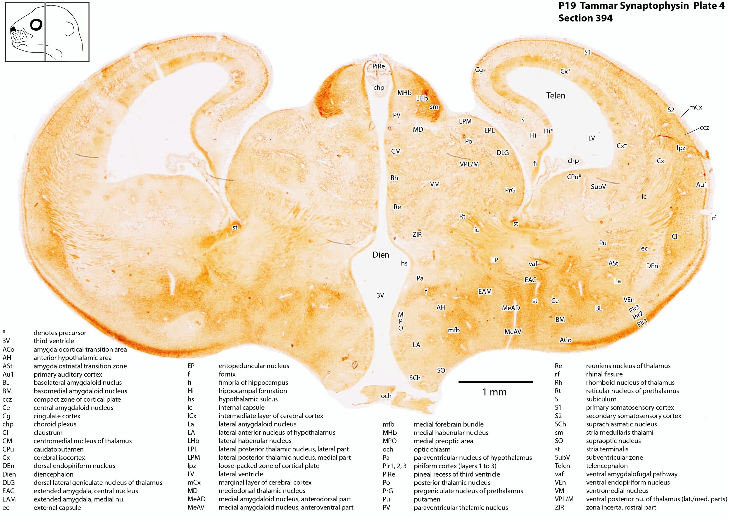
Immunoreactivity for synaptophysin is less focal in the P19 tammar brain than in the P0 brain, but most immunoreactivity is still found in the tegmentum of the myelencephalon of the brainstem (plate 8).
In the forebrain, most immunoreactivity is found in components of the olfactory pathways, including the external plexiform layer of the olfactory bulb (EPl, plate 1), the lateral olfactory tract (lo, plates 1 to 3), and the superficial layer of the piriform cortex (Pir1, plates 3, 4).
Other areas of strong synaptophysin immunoreactivity include a series of fibre pathways, such as the fornix (f, plate 3), stria medullaris sthalami (sm, plate 4) external capsule (ec, plate 3), internal capsule (ic, plate 3) and the fasciculus aberrrans (fa, the commissural pathway from the internal capsule, plate 3, Elliott Smith 1902).
Strong immunoreactivity in the brainstem is found in the cerebellum and its outflow, the superior cerebellar peduncle (scp, plate 8), and in the trigeminal sensory nuclear complex (e.g. Sp5O, plate 8).
Reference
Elliott Smith G (1902) On a peculiarity of the cerebral commissures in certain Marsupialia, not hitherto recognised as a distinctive feature of the Diprotodontia. Proc Roy Soc Lond 70: 226-231.
Tarsa L, Goda Y (2002) Synaptophysin regulates activity-dependent synapse formation in cultured hippocampal neurons. Proc Natl Acad Sci 99: 1012-1016.