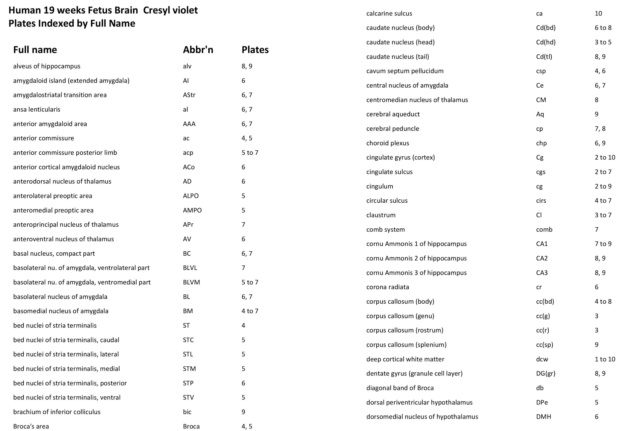
Atlas of the Fore- and Midbrain of a 19 week Human Fetus
Introduction
This specimen is of a 19 week post-conception human brain. The cerebellum, pons and medulla had been removed, so frontal or coronal images shown here are only of the fore- and midbrain. The human brain at this stage has all major components formed, but is still in an intense period of small neuron and macroglia generation.
Methods
The brain was fixed in 10% buffered formalin. The left half of the brain was coronally sectioned at a thickness of 50 µm with a freezing microtome. Every 10th section was mounted onto subbed slides and stained with cresyl violet before being coverslipped. The sections were photographed with a Canon 4000D and an EFS 60 mm FL macrolens. Images were photomontaged with Adobe Photoshop 2023 and labelled with Adobe Illustrator 2023.
General Features
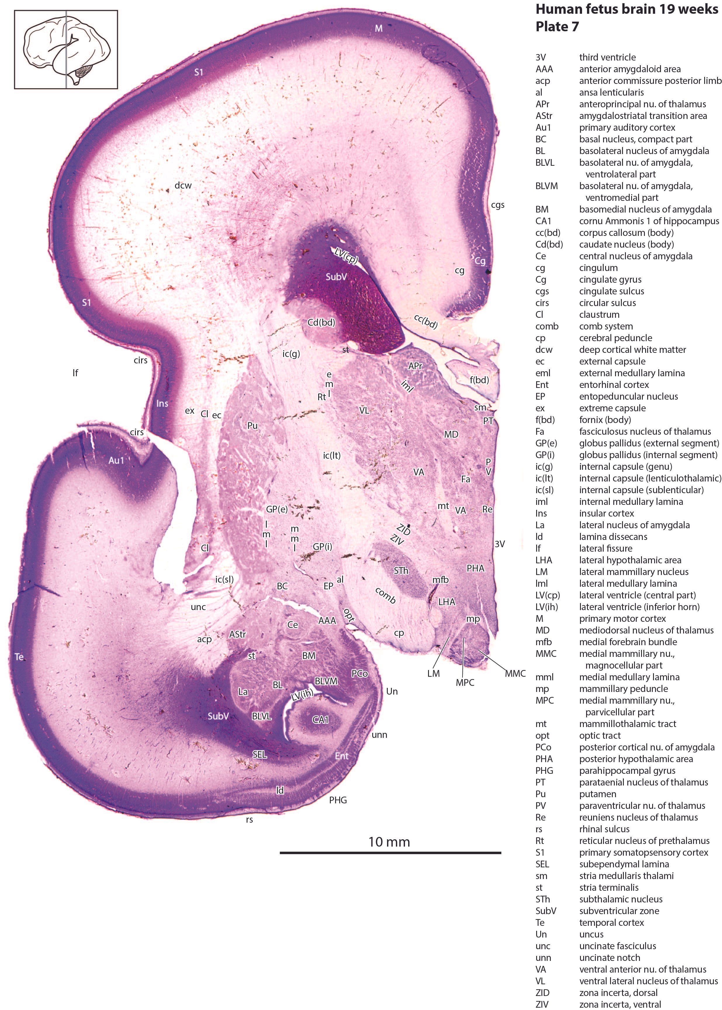
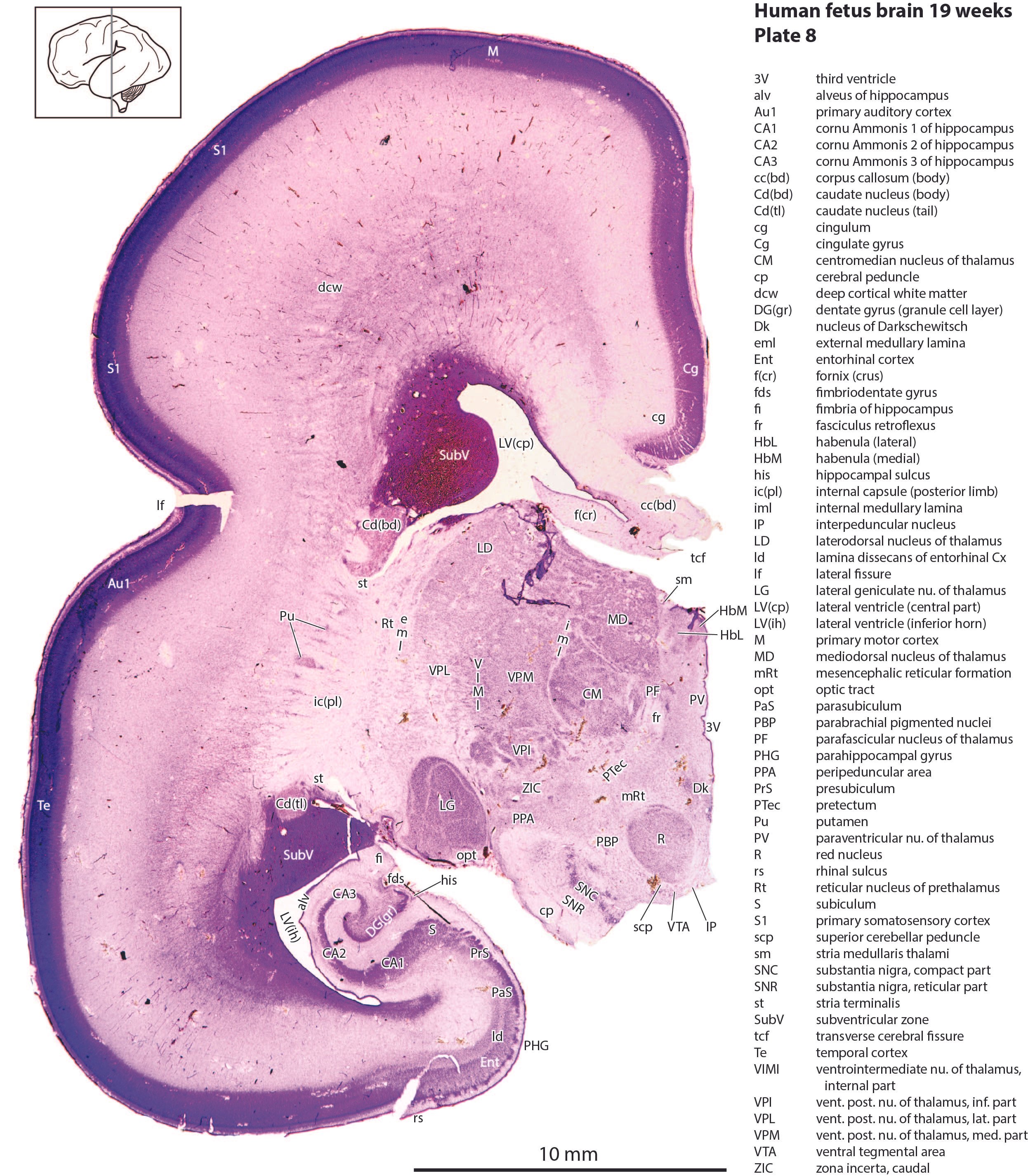
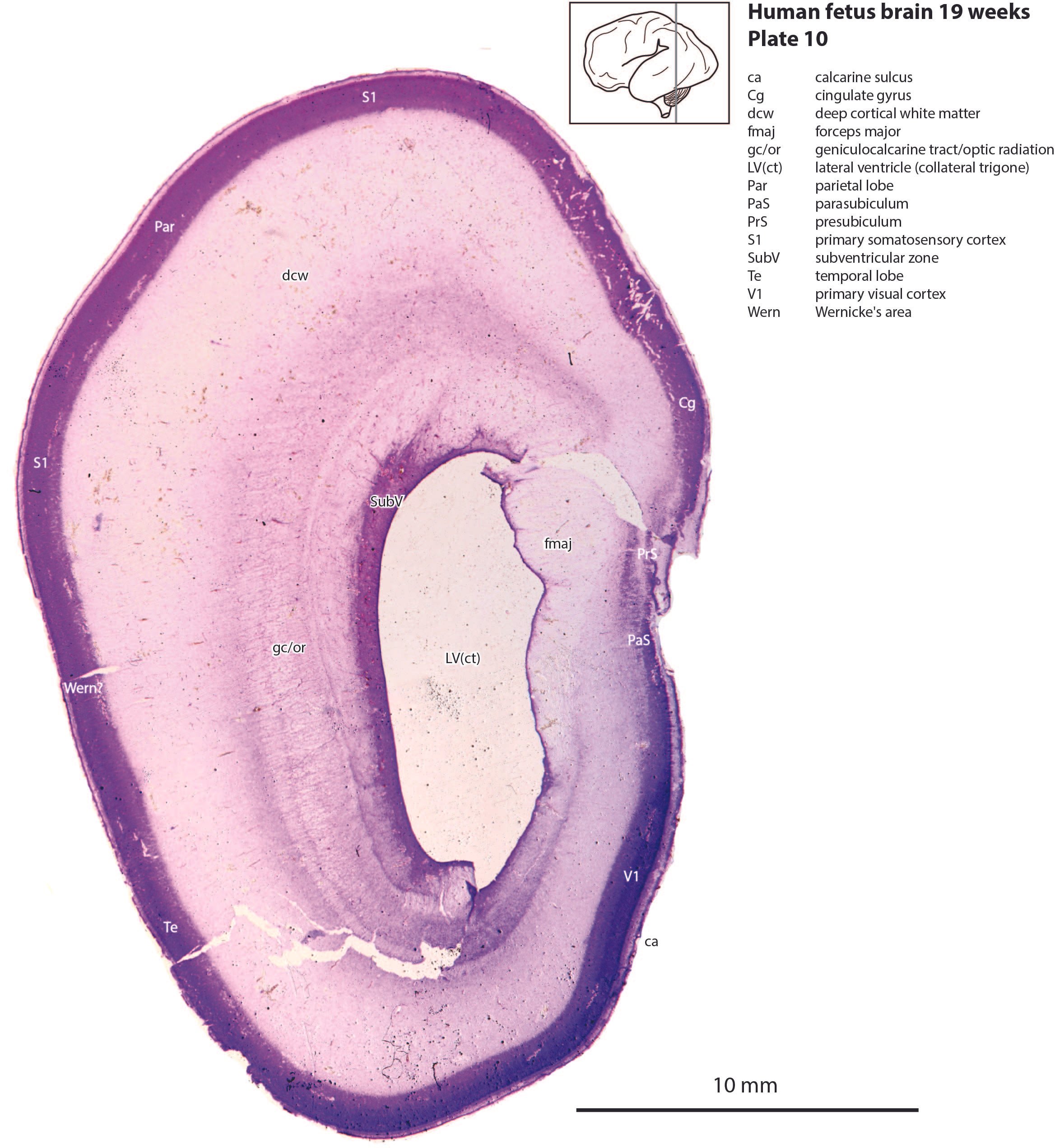
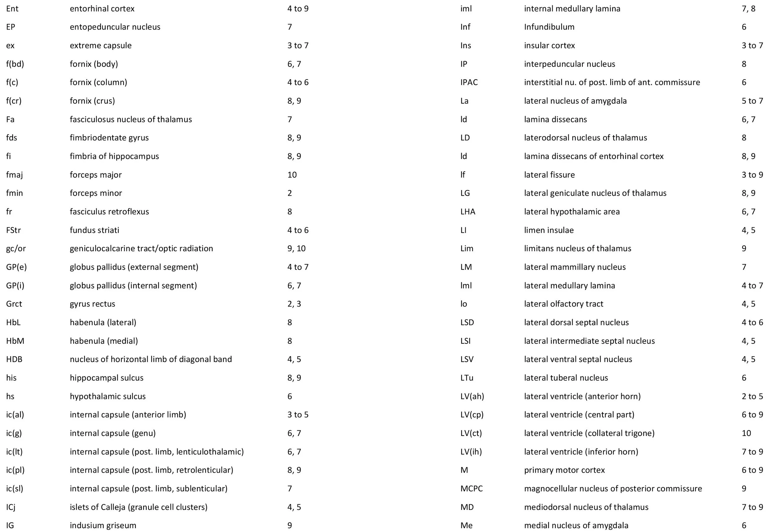
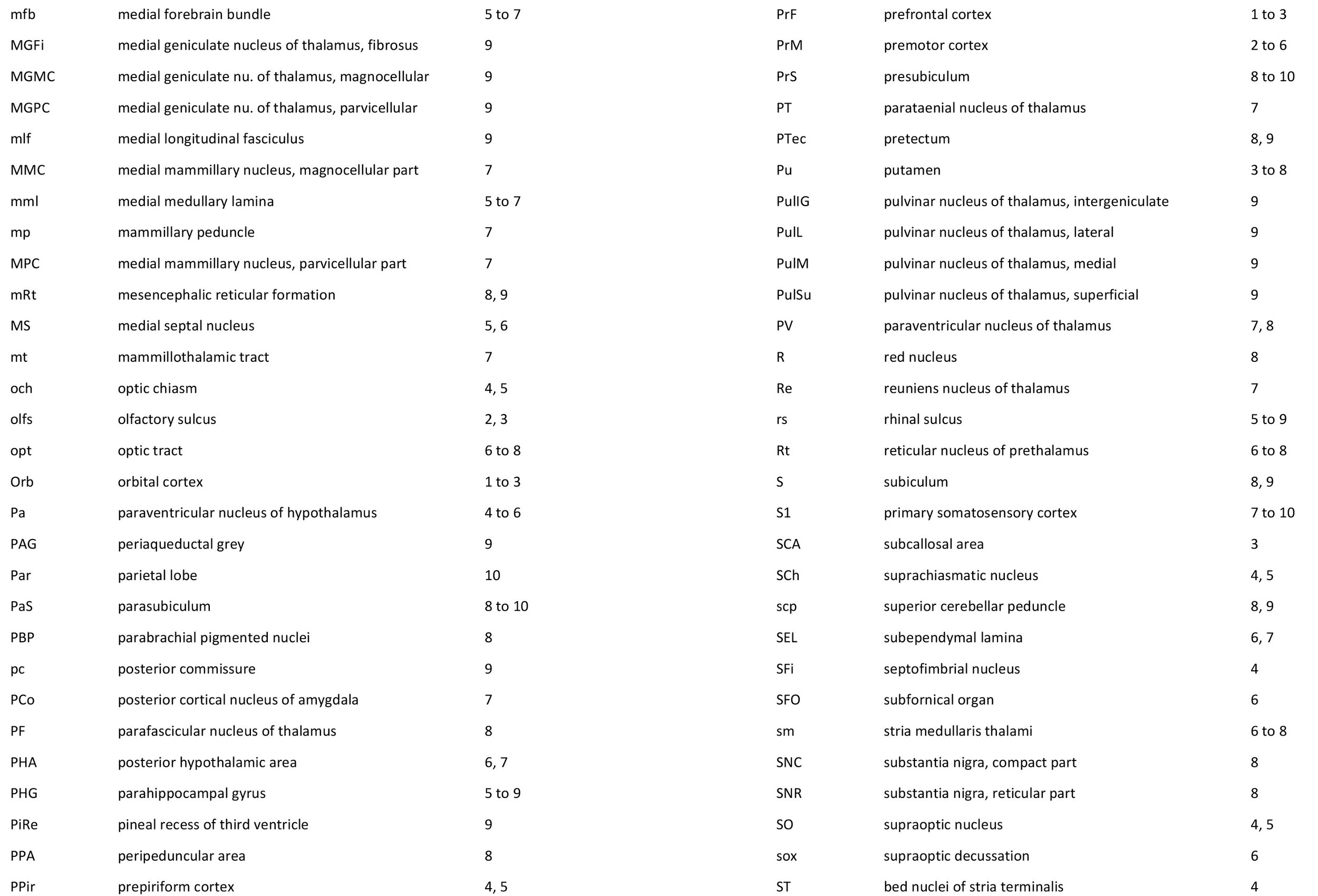
The surface of the brain shows a few fissures and sulci. The most prominent of these is the lateral fissure (lf in plates 3 to 9) which separates the frontal and parietal lobes from the temporal lobe. The insula cortical region lies in the depths of the lateral fissure and is encircled by a circular sulcus (cirs in plates 4 to 7), which meets the ventral brain surface at the limen insulae (LI in plates 4, 5). Other smaller and subtle sulci include the olfactory sulcus (olfs in plates 2, 3) which houses the olfactory bulb and tract during life, the cingulate sulcus (cgs in plates 2 to 7), and the calcarine sulcus (ca in plate 10) where the primary visual cortex will form. The fimbriodentate sulcus (fds in plates 8, 9) and hippocampal sulcus (his in plates 8, 9) are associated with the hippocampal formation, while the rhinal sulcus (rs in plates 5 to 9) marks the lateral boundary of the parahippocampal gyrus.
Telencephalon
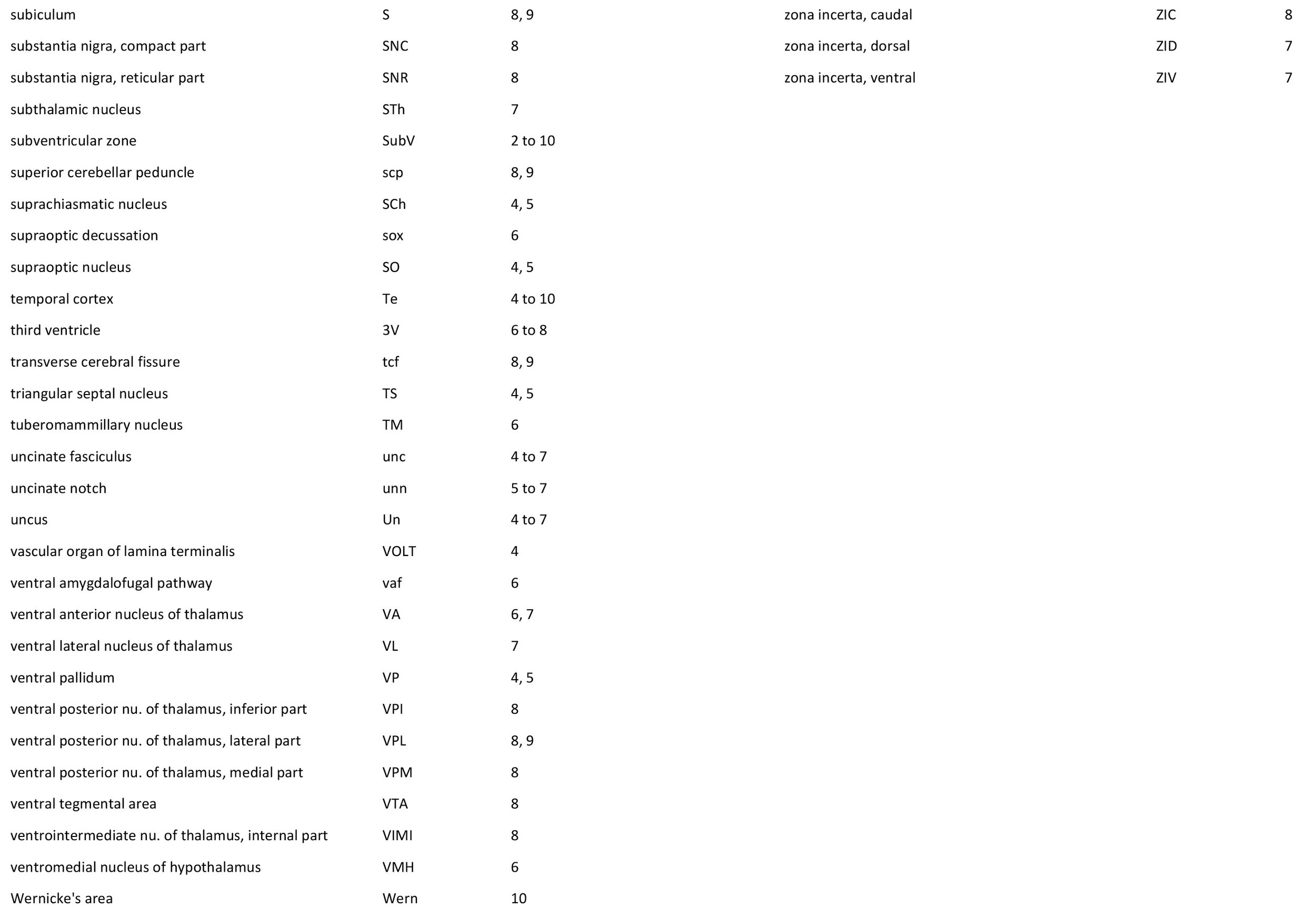
The bulk of the telencephalon is formed by the developing pallium (cortex), underlying white matter and transitional zones for migrating neurons and glia, and the deeper basal ganglia (caudate, putamen, pallidum). The lateral ventricle is surrounded by a thick subventricular zone (SubV in plate 2 to 10), which at this stage, generates small neurons and glia.
Lamination of the cerebral cortex is still indistinct. The early formed deeper layers (layers 5 and 6) have emerged in the lower levels of the cortex, but more recently generated small neurons of the superficial layers (granular and supragranular layers; layers 4, 3 and 2) are still clumped in a dense cortical plate. There are also many neurons and macroglia in the process of migrating through the embryonic white matter layers (transitional zones) to the cortical plate.
All the major elements of the basal ganglia (striatum and pallidum) are in position, but would still be receiving migrating microneurons from the subventricular zone. The caudate nucleus has a head, body and tail (Cd in plates 3 to 9). The lentiform nucleus includes the putamen (Pu in plates 3 to 8) and external and internal segments of the globus pallidus (plates 4 to 7).
All components of the internal capsule can be seen even at this stage. These include the anterior limb ic(al) in plates 3 to 5, genu ic(g) in plates 6, 7 and lenticulothalamic, sublenticular and retrolenticular parts of the posterior limb of internal capsule (see plates 6 to 9).
Most of the components of the amygdala can be distinguished (BM, BL, Ce, Me; see plates 4 to 7).
Diencephalon
In modern conceptions the diencephalon is divided caudorostrally into pretectum, thalamus and prethalamus, derived respectively from prosomeres 1, 2, and 3. Very little detail is visible in the pretectum (PTec in plates 8, 9), but the thalamus and prethalamus show many of the major adult nuclei. In particular note components of the VP group (for somatosensation; e.g. VPM in plate 8, VPL in plates 8, 9), the lateral geniculate nucleus (LG in plates 8, 9) for vision, and the MG components (for audition; MGFi, MGMC, MGPC in plate 9). The prethalamus is represented by the reticular thalamic nucleus (Rt in plates 6 to 8), subthalamic nucleus (STh in plate 7) and the zona incerta (ZIC, ZID, ZIV in plates 7, 8).
Midbrain
Only the most rostral parts of the midbrain are present, including the red nucleus (R in plate 8), the substantia nigra (SNC, SNR in plate 8) and the mesencephalic reticular formation (mRt in plates 8, 9).