Atlas of the Brain of a Grey Whale Fetus (Eschrichtius robustus)
Introduction
The grey whale is a bottom-feeding baleen whale reaching up to 15 m in length and 20 tonnes in weight. The modern population is mainly found in the eastern North Pacific, feeding mainly on benthic crustaceans. Grey whales migrate between polar latitudes (Alaska) and warmer waters (e.g. Baja California) where the females give birth (Rice et al., 1984). Life expectancy is up to 70 years.
Gestation period is about 400 days and calves can be up to 4.6 m in length at birth (Agbayani et al., 2020).
Materials and Methods
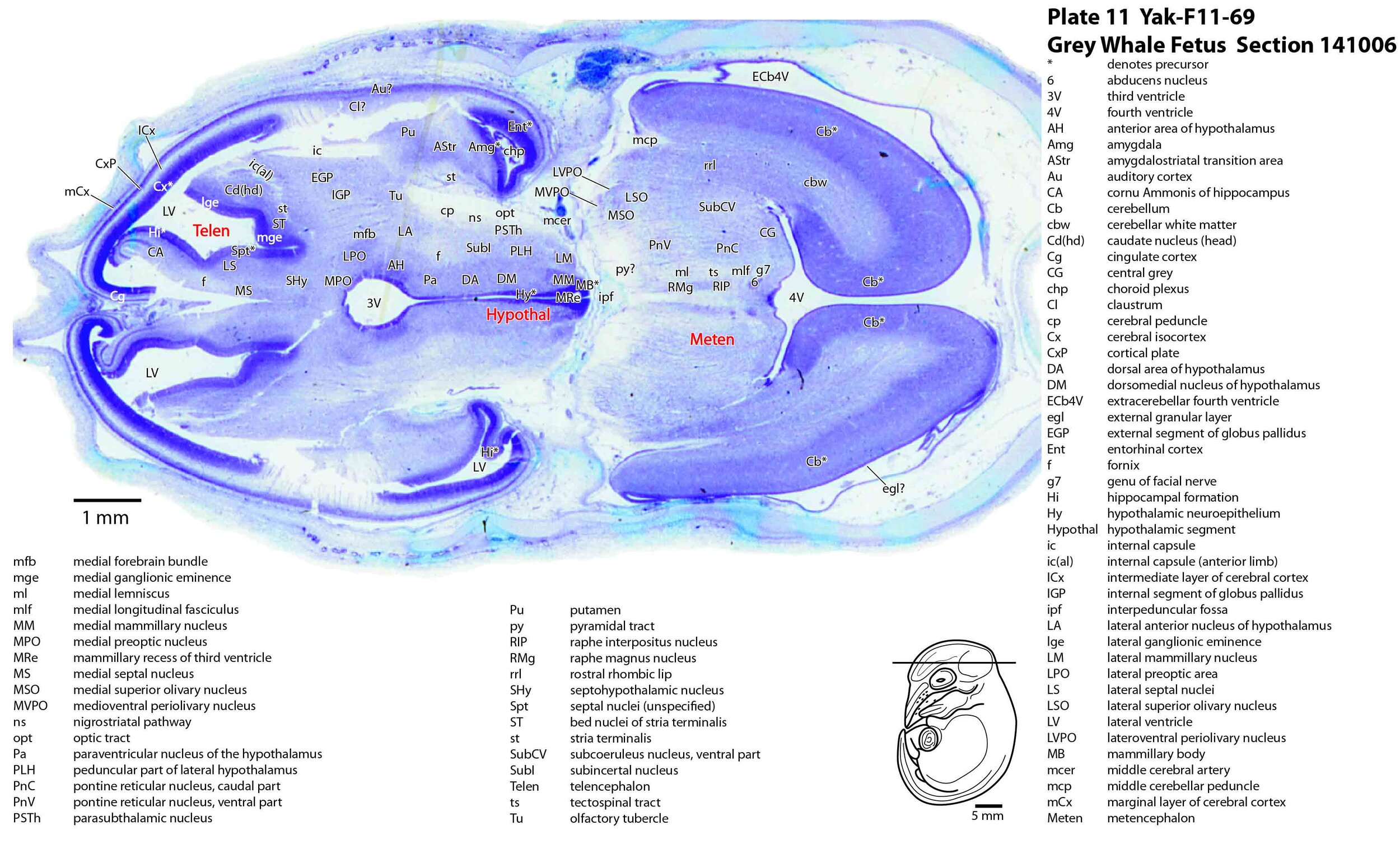
The fetal head and brain depicted here is Yak-F11-69 from the Yakovlev collection and the Nissl-stained sections are held at the National Museum for Health and Medicine in Silver Springs, Maryland, USA. The brain is approximately 17 mm long (rostral to caudal) in the dehydrated state and would have been 24 mm long in an undehydrated state. The crown-rump length of the intact fetus would have been approximately 32 mm. The fetus shown here would be only approximately 1.5 to 2.0 % of the length of a newborn, so there would be a prolonged period of intrauterine growth after the stage of this fetus.
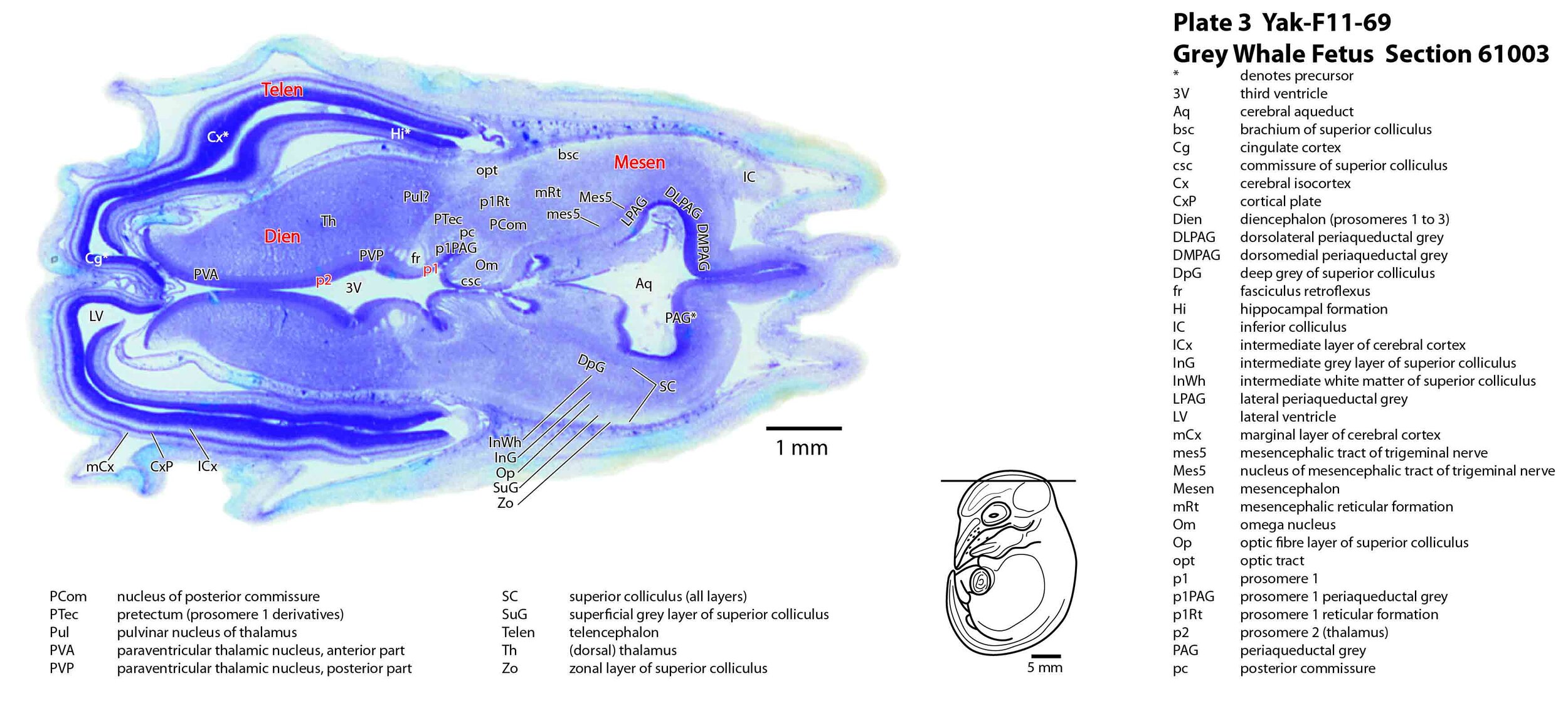
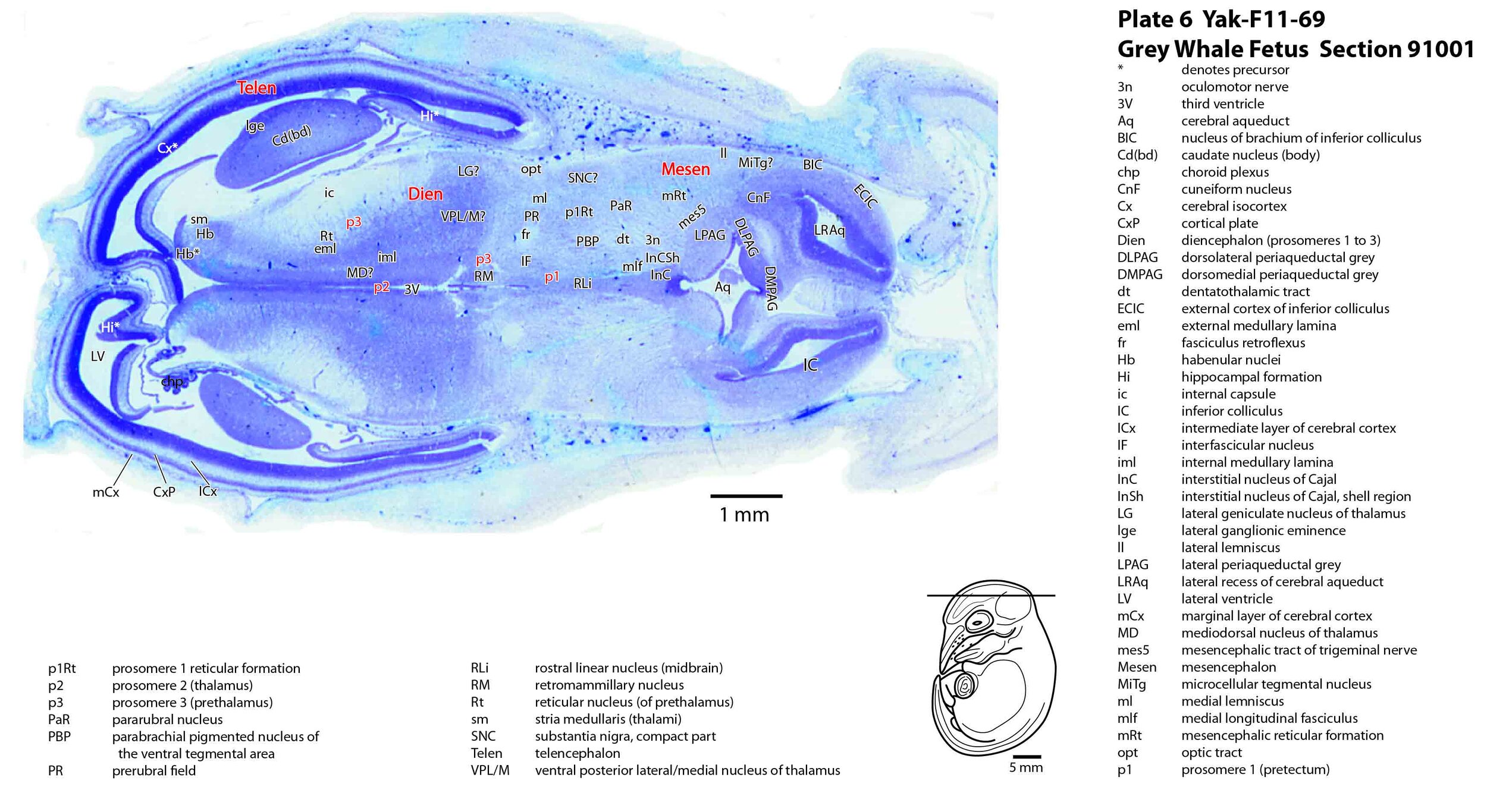
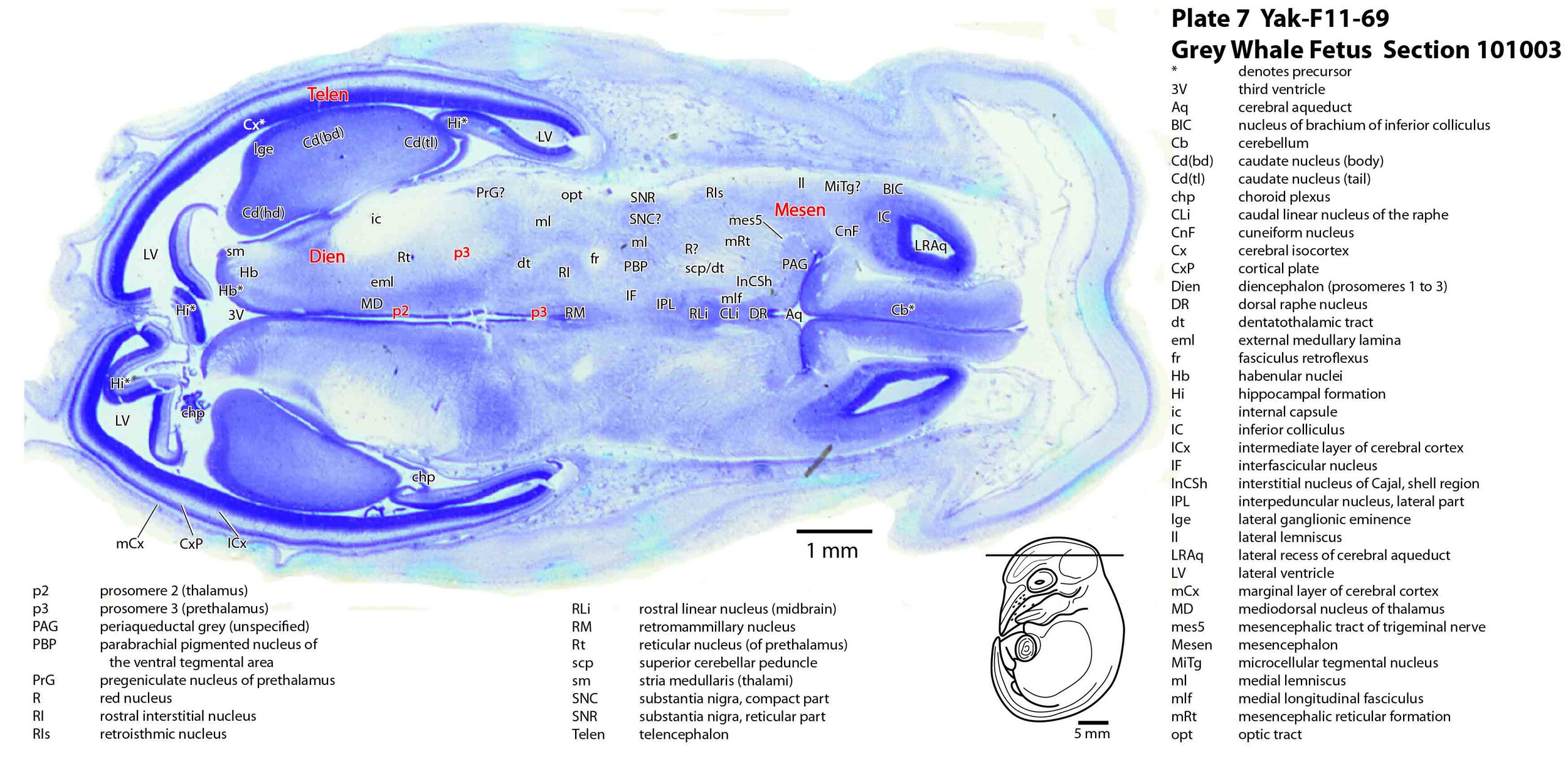
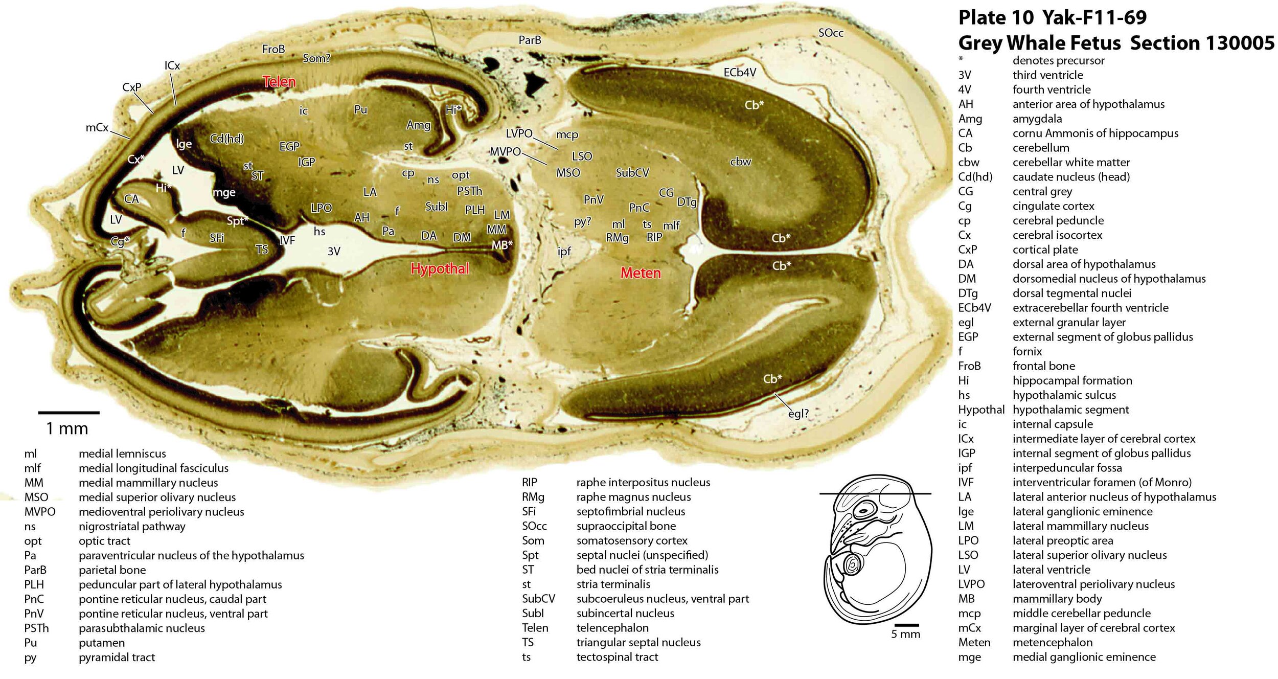
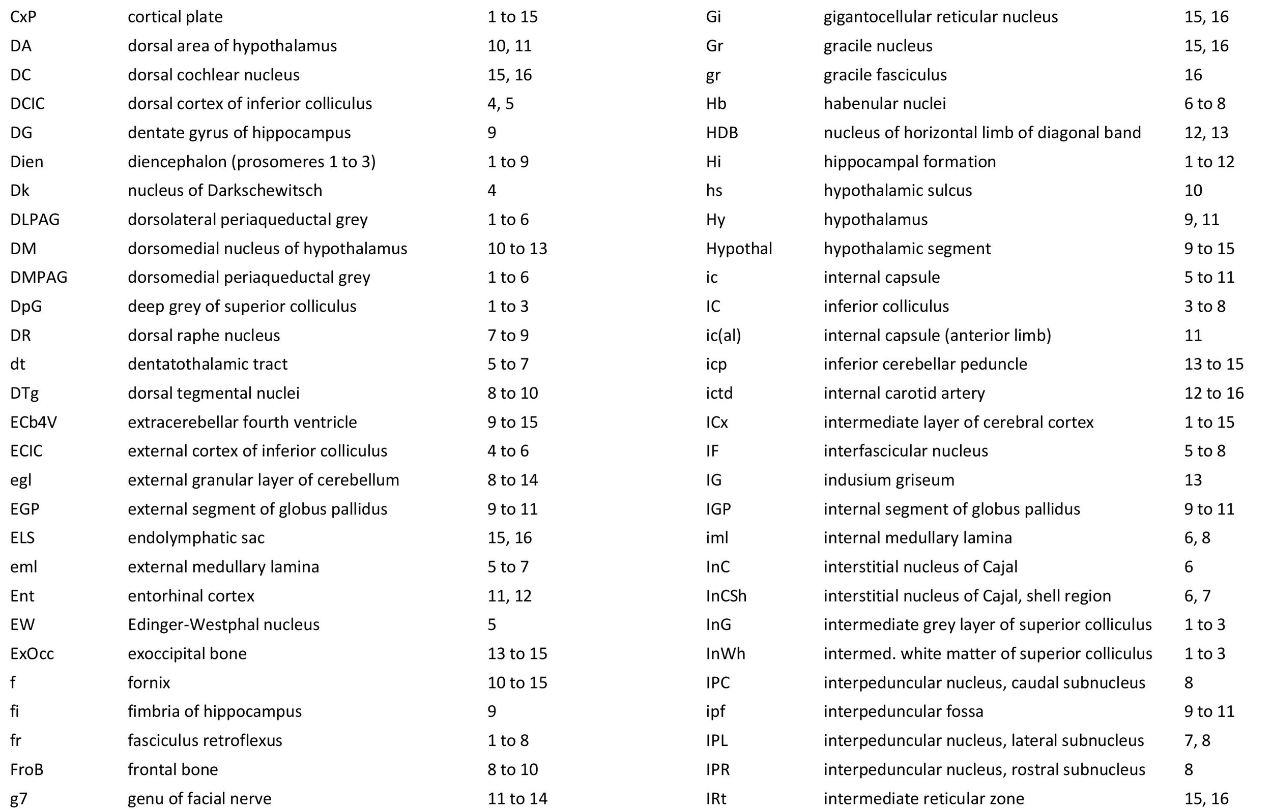
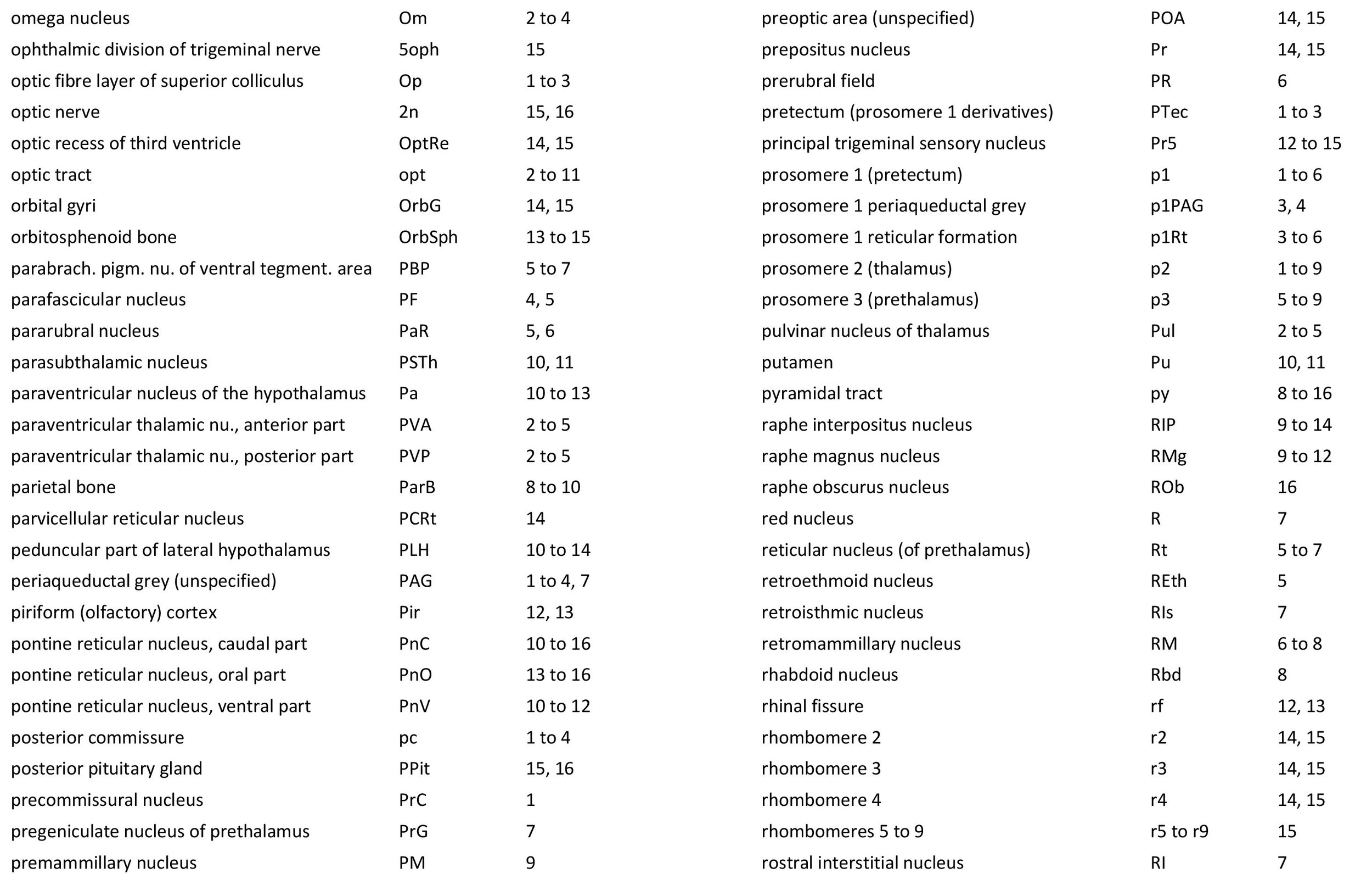
Selected horizontal plane sections in a regular sequence (please see section numbers on plates) were scanned with the aid of an Epson 11000. The images were cropped and adjusted for optimal contrast levels with Adobe Photoshop(2021), before 16 sections were placed into 20 Adobe Illustrator (2021) files for mapping. Only one side of the brain (right side of the section) has been labelled to leave the other side unobscured. Regions of the brain were labelled using terminology adapted from that used in the Paxinos and Watson system of brain atlases. The section number is given in the top right-hand corner of the plate. A small line-diagram finder image in each atlas plate shows the position of the horizontal section (grey line) within the fetal brain. No image of the original intact specimen was available, so the finder image is based on a humpback whale fetus of similar size and age (Hampe et al., 2015). Finished atlas plate files were exported to jpg for publishing.
Description of Specimen
Features of the brain will be described in a rostrocaudal sequence starting with the telencephalon and finishing with the myelencephalon.
The cerebral isocortex (Cx) is smooth (plates 1 to 15), with a thin neuroepithelium, a thin but distinct intermediate layer (ICx), and an early cortical plate (CxP). There is no differentiation of cortical regions. The archicortex has begun to infold to produce an hippocampal primordium (Hi) with cornu Ammonis (CA; plates 9 to 12) and a very small dentate gyrus (DG; plate 9).
The lateral ventricle is dominated by a large lateral ganglionic eminence (lge; plates 6 to 13), which is in the process of generating the (neo)striatum (caudate - Cd and putamen - Pu); and a smaller medial ganglionic eminence (mge; plates 8 to 13), adjacent to the bed nuclei of the stria terminalis.
The diencephalon consists of three rostrocaudal prosomere segments (from caudal to rostral; p1 – pretectum; p2 – thalamus, previously dorsal thalamus; and p3 – prethalamus, previously ventral thalamus; see plate 5). Prosomere p2 is separated from p3 by a zona limitans intrathalamica (zli; plates 8, 9), while the descending fasciculus retroflexus (fr; plates 1 to 8) marks the approximate boundary between p1 and p2. Differentiation of individual nuclei within all three diencephalic segments has begun, but is far from complete.
There is a large internal capsule (ic; plates 5 to 11) between the thalamus and striatum, which is probably early thalamocortical axons and striatal connections because the white matter beneath the isocortical plate is still very thin at this stage, suggesting that very few corticostriatal, corticothalamic and/or corticocortical connections have been established at this age.
There is no sign yet of a corpus callosum, hippocampal commissure, or anterior commissure.
The brainstem (midbrain to medulla oblongata) is the most differentiated structure of the brain at this stage. Lamination of the superior colliculus of the mesencephalon (midbrain) has begun to appear (plates 1 to 3) and the auditory inferior colliculus (plates 3 to 8) is large and expanding from neurogenesis in the neuroepithelial walls of paired lateral recesses of the cerebral aqueduct (LRAq; plates 5 to 7).
Major efferent nuclei of the brainstem (motor trigeminal – 5; abducens – 6; dorsal motor nucleus of vagus nerve – 10; hypoglossal – 12; see plates 13 to 16) show as darker structures due to accumulated Nissl substance. Some major pathways are visible (medial lemniscus, medial longitudinal fasciculus, superior cerebellar peduncle).
The developing cerebellum consists of paired everted wings extending from the rostral rhombic lip (rrl; plates 11 to 15) and surrounded by an extracerebellar part of the fourth ventricle. At this stage the midline cerebellar vermis is unfused (e.g. plates 11 and 12) and internal detail within the cerebellum (e.g. deep cerebellar nuclei, Purkinje cell lamination) is difficult to see, although a thin external granular (or germinal) layer (egl) for production of cerebellar microneurons coats the outer surface of the cerebellar primordium.
Acknowledgements
I would like to thank Elizabeth Lockett of the National Museum of Health and Medicine in Silver Springs, Maryland, USA for the opportunity to study the marine mammal brains in the NMHM collection and for use of the Epson 11000scanner.
References
Agbayani S, Fortune SME, Trits AW (2020) Growth and development of North Pacific gray whales (Eschrichtius robustus) J Mammal 101: 742-754.
Hampe O, Franke H, Hipsley CA, Kardjilov N, Müller J (2015) Prenatal cranial ossification of the Humpback Whale (Megaptera novaeangliae). J Morphol. 276: 564-582.
Rice DW, Wolman AA, Braham HW (1984) The gray whale, Escherichtius robustus. Marine Fisheries Review 46: 7-14.