Atlas of the Brain of a Harbour Seal (Phoca vitulina)
Introduction
The harbour seal (Phoca vitulina) is also known as the common seal and is found along arctic and temperate coastlines in the northern hemisphere. Adults can reach 1.85 m in length and over 180 kg. True seals belong to the Order Carnivora and this is reflected in the brain structure, which is similar to felids and canids (see below).
Materials and Methods
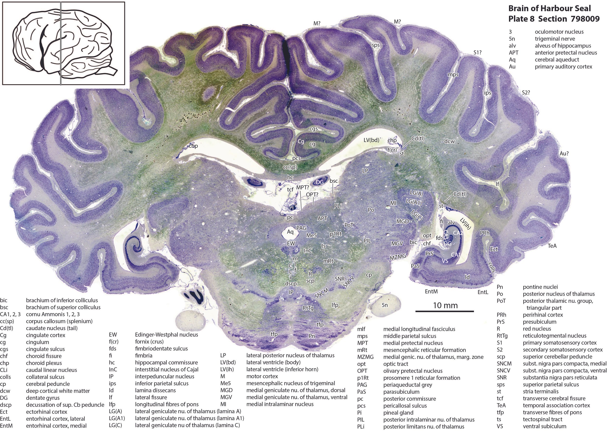
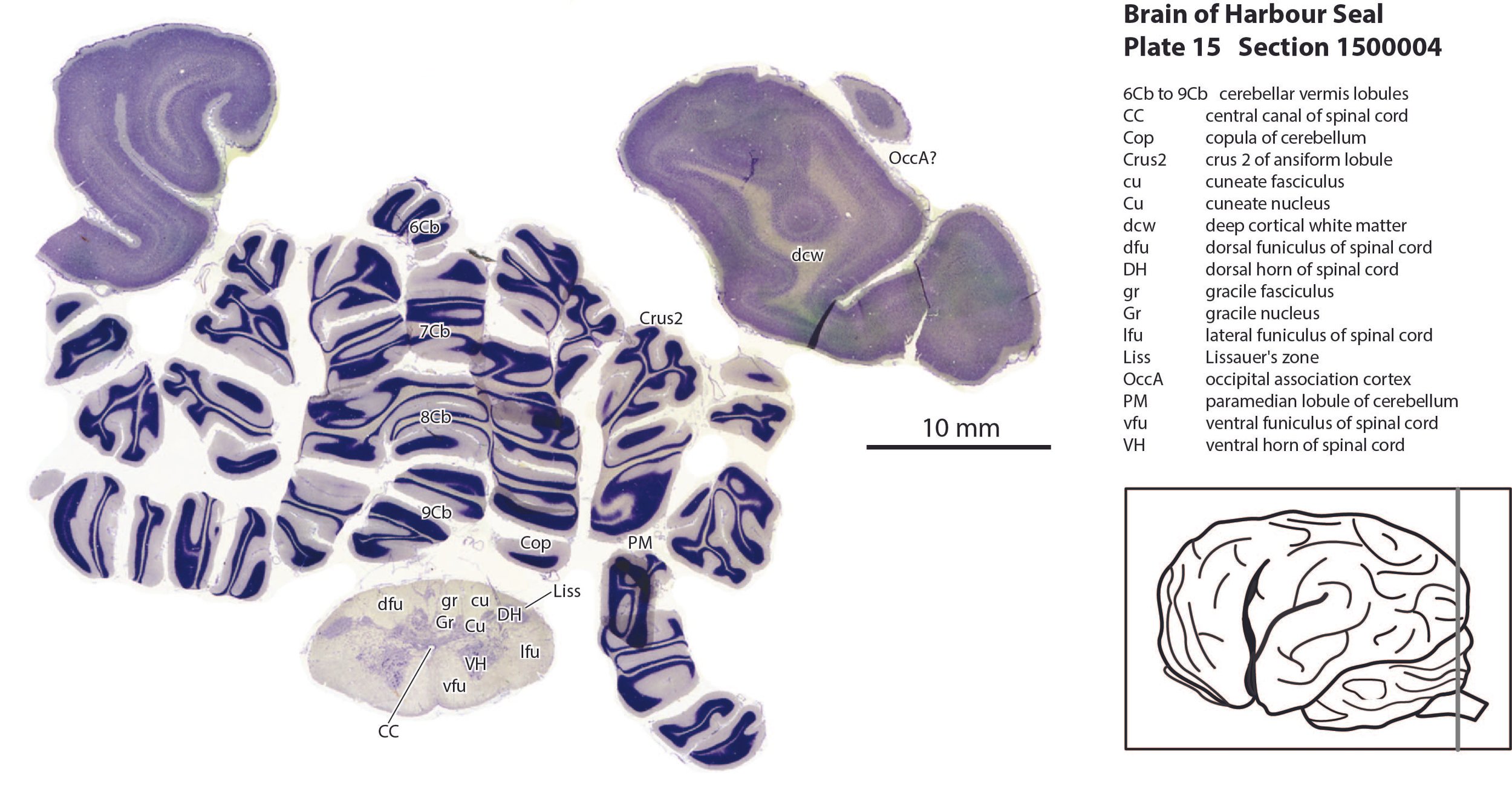
This specimen is part of the Comparative Mammalian Brain Collection (Wally Welker, John Irwin Johnson, Adrianne Noe) that can be seen at brainmuseum.org. The physical sections are held at the National Museum for Health and Medicine (NMHM) at Silver Springs Maryland, USA. Details of tissue preparation and histology can be found at brainmuseum.org. Selected frontal plane sections in a regular sequence (please see section numbers on plates) were scanned with the aid of an Epson 11000. The images were cropped and adjusted for optimal contrast levels with Adobe Photoshop (2023), before 16 sections were placed into 15 Adobe Illustrator(2023) files for mapping. Only one side of the brain (right side of the section) has been labelled to leave the other side unobscured. Regions of the brain were labelled using terminology adapted from that used in the Paxinos and Watson system of brain atlases. The section number is given in the top right-hand corner of the plate. A small line-diagram finder image in each atlas plate shows the position of the horizontal section (grey line) within the fetal brain. An image of the original intact specimen was available at brainmuseum.org. Finished atlas plate files were exported to jpg for publishing.
Description of Specimen
The cerebral cortex of the brain is highly gyrified (gyrification index of 2.51 compared to 1.88 and 1.56 for the domestic dog and cat, respectively; Ashwell and Gurovich, 2019), so a system of prominent gyri and deep sulci can be identified. The most prominent groove on the cortical surface is the lateral fissure (lf in plates 4 to 9) separating the frontal and temporal lobes.
The olfactory bulb (plate 1) is relatively small and housed in a dorsally turning olfactory sulcus/rhinal fissure between the gyrus rectus and orbital gyri. The anterior olfactory area (AO in plates 1, 2) is particularly small and located along the olfactory tract. Piriform cortex (Pir in plates 3 to 6) is located on the ventral surface of the temporal lobe.
The hippocampal formation is mainly located in the temporal lobe (plates 7, 8), but also has an extension into the dorsal part of the brain ventral to the corpus callosum (see DG in plate 8).
The thalamic visual relay nucleus (lateral geniculate or LG in plates 8 and 9) and the adjacent medial intralaminar nucleus (MI in plate 8) are very similar in structure to those in the domestic cat, with prominent A, A1 and C laminae in the LG. Based on similarities to the domestic cat (see review in Stacy and Van Hooser, 2002), it is likely that the contralateral eye projects to the A, C and C2 laminae of the LG and the ipsilateral eye projects to the A1 and C1 laminae.
Acknowledgements
I would like to thank Elizabeth Lockett of the National Museum of Health and Medicine in Silver Springs, Maryland, USA for the opportunity to study the marine mammal brains in the NMHM collection and for use of the Epson 11000 scanner.
References
Ashwell KWS, Gurovich Y (2019) Quantitative analysis of forebrain pallial morphology in monotremes and comparison with that in therians. Zoology (Jena). 134: 38-57.
Stacy AK, Van Hooser SD (2022) Development of functional properties in the early visual system: new appreciations of the roles of the lateral geniculate nucleus. Current Topics in Behavioral Neuroscience. 53.