Atlas of the Brain of a 5 week Human Embryo, Carnegie #1006
Introduction
This specimen is at a stage when all five secondary brain vesicles have appeared, although the evagination of the telencephalic vesicles is incomplete. The bulk of brain tissue is made up of proliferative compartments (neuroepithelium), with only very small populations of (post-mitotic) neurons in the rhombencephalic tegmentum. Segmentation is visible in the forebrain into prosomeres (diencephalon), hypothalamic segments (hy1, hy2, hyat), pallium or protocortex and ganglionic eminences (telencephalon).
Methods
The specimen illustrated here is Carnegie #1006, held at the National Museum for Health and Medicine (NMHM) in Silver Springs, Maryland. Likely crown-rump length is between 7 and 9 mm. Estimated post-conception age is approximately 35 days.
The specimen was acquired in 1914, fixed in formol, embedded in paraffin, sectioned coronally at a thickness of 20 µm, and the sections stained with hematoxylin and eosin or “G” (O’Rahilly and Müller, 1987).
Raw images were collected by Ken Ashwell at the NMHM in 2014, using a Nikon E800 microscope with a Ludl Biopoint automated stage and a MAC 2000 controller. Photomicrographs were taken with Micropublisher V5, with ImagePro Plus software from Media Cybernetics.
All images were calibrated by photographing a scale bar at the same magnification. Images were photomontaged and optimised for contrast using Adobe Photoshop 2023 before being placed in Adobe Illustrator 2023 for delineation and labelling. Developmental regions (i.e. neuroepithelium) destined to give rise to adult structures have been denoted by the adult structure’s name with an asterisk (e.g. Cx* denotes the developmental field of the cerebral isocortex).
A line diagram of a stage 15 human embryo has been used for the finder illustration indicating section position.
Notes on the specimen
General observations
The only part of the brain where neurogenesis has commenced is the rhombencephalon (brainstem) where a few nuclear groups have begun to appear in the thin tegmentum. The forebrain shows prosomeric divisions of the neural tube, but no significant post-mitotic populations. The cerebral isocortex (Cx* in plates 2, 3) is rudimentary and consists of only proliferative populations. The cerebellum is represented only by the brim of the rostral rhombic lip.
Central nervous system
The telencephalic vesicles have partially evaginated, but the lateral ventricles are connected to the third ventricle by a wide interventricular foramen. The telencephalic wall consists only of a proliferative compartment (ventricular germinal zone) about 150 to 200 µm thick. There is no preplate visible in the pallium (cerebral cortex).
The hypothalamus is divided into peduncular (hy1), tuberal/terminal (hy2), and acroterminal (hyat) proliferative components (variously in plates 2 to 5). There is no significant formation of hypothalamic nuclei external to the proliferative compartments. There is a prominent optic stalk (os in plate 4) connecting the eye cup with the hypothalamus.
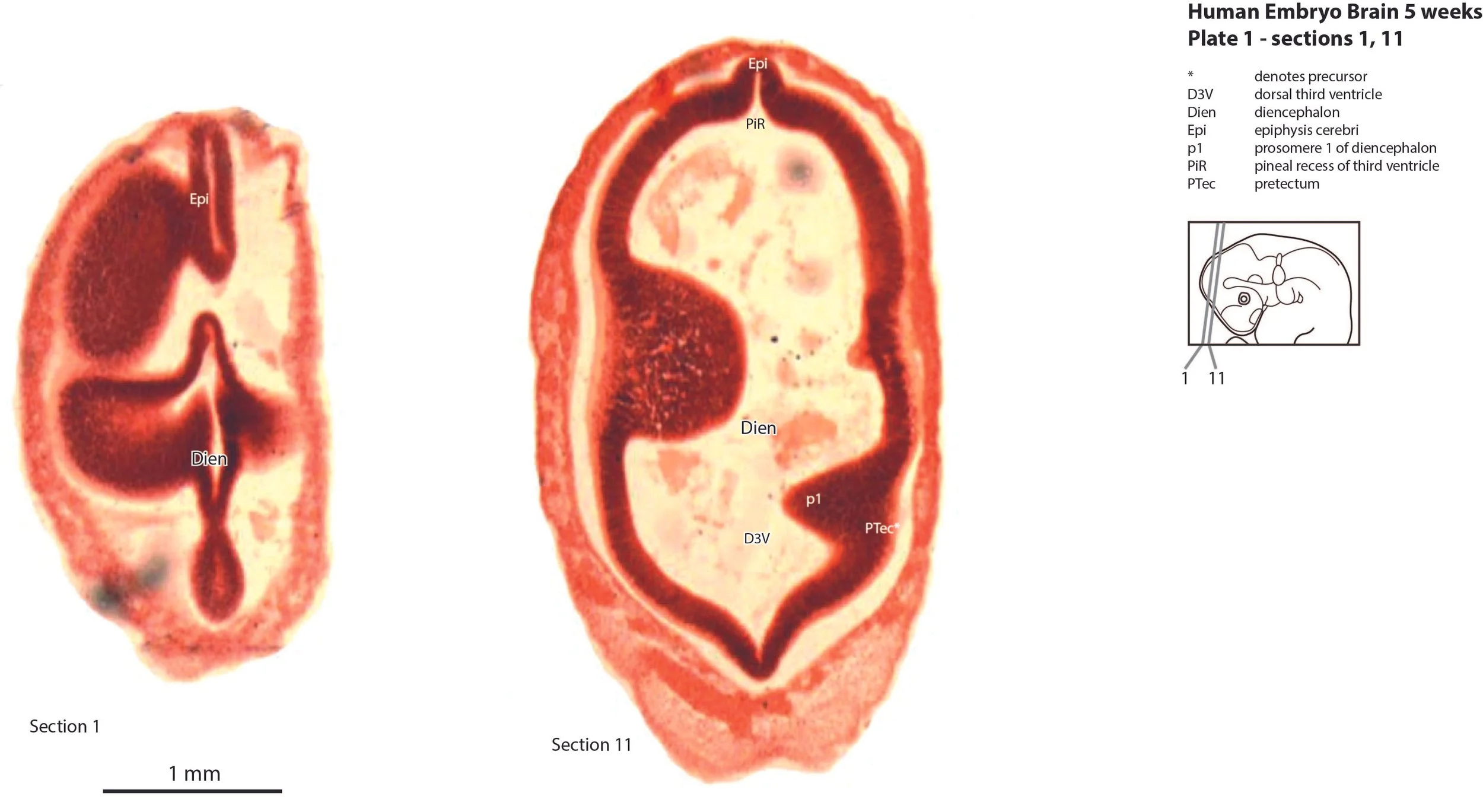
The diencephalon has the typical prosomeric structure. Prosomeres 1 (pretectum), 2 (dorsal thalamus), and 3 (prethalamus) can be identified as proliferative zones (see plates 1 to 3), but postmitotic populations are negligible, particularly in the dorsal thalamus (Th in plates 2,3).
The mesencephalon (Mesen in plate 2) has a tubular structure with a broad cerebral aqueduct (Aq in plate 2), where most of the neuraxis wall is made up of the proliferative neuroepithelium, and very few postmitotic neurons. The mesencephalic tegmentum is very poorly developed.
The cerebellum (Cb in plate 4) consists solely of a thin proliferative compartment rim extending from the rostral rhombic lip. There are no neuronal populations present.
The brainstem is the most differentiated part of the brain. There is a thin trigeminal sensory nuclear complex (Pr5, Sp5O, Sp5I; variously in plates 5 to 7) in the lateral tegmentum of the brainstem. There is also a thin medial magnocellular reticular formation (Gi; plates 6 to 7).
Sense organs and peripheral nervous system
Some cranial nerves can be identified. Cranial nerve 2 (optic) is not developed, but an optic stalk (os in plate 4) extends from the hypothalamus to the eye cup. The eye muscle nerve 3n (oculomotor) can be traced from the brainstem to the orbit (3n in plates 2 to 4). 4n and 6n are not visible, but must be present. The trigeminal nerve (5n in plate 5) runs from the trigeminal ganglion (5Gn in plates 4, 5) to the brainstem. The facioacoustic ganglion complex is breaking up into its component geniculate (Gen in plate 7), vestibular (VeGn in plate 7) and cochlear (CGn in plate 7) subunits. The otocyst is still very simple in structure and its main feature is a primitive cochlear duct (CD in plate 6). The hypoglossal nerve is not visible, but the hypoglossal nucleus is apparent (12 in plate 7).
Acknowledgements
I would like to thank Dr Elizabeth Lockett of the NMHM for access to the collection and for all her kind help during the work.
References
O’Rahilly R, Müller F (1987) Developmental stages in human embryos. Carnegie Institution of Washington 63T.