Atlas of the Brain of a 3.5 week Human Embryo, Carnegie #164
Introduction
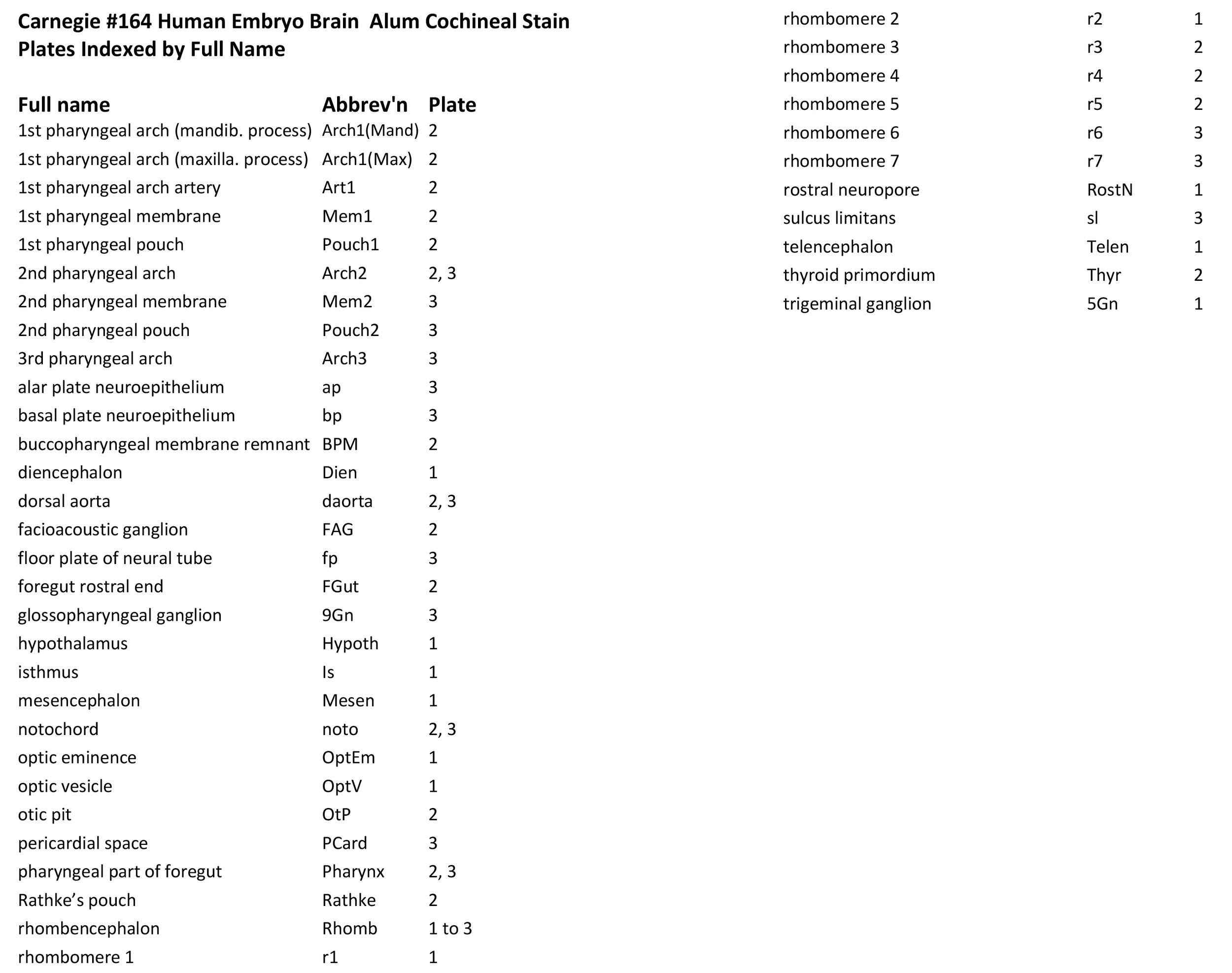
This specimen is at a stage (Stage 11) when the rostral or cephalic neuropore (RostN in plate 1) of the neural tube has just closed, but the caudal neuropore remains open. The optic vesicle is forming from the hypothalamus, but all levels of the neural tube are primitive and the neuroepithelium thin.
Methods
The specimen illustrated here is Carnegie #164, held at the National Museum for Health and Medicine (NMHM) in Silver Springs, Maryland. Crown-rump length is 3.5 mm. Estimated post-conception age is approximately 24 days (O’Rahilly and Müller, 1987), although in another publication O’Rahilly and Müller (2006) have listed stage 11 embryos as 30 postfertilization days.
The specimen was acquired in 1913, fixed in formol, embedded in paraffin, sectioned transversely at a thickness of 20 µm, and the sections stained with alum cochineal (O’Rahilly and Müller, 1987).
Raw images were collected by Ken Ashwell at the NMHM in 2014, using a Nikon E800 microscope with a Ludl Biopoint automated stage and a MAC 2000 controller. Photomicrographs were taken with Micropublisher V5, with ImagePro Plus software from Media Cybernetics.
All images were calibrated by photographing a scale bar at the same magnification. Images were photomontaged and optimised for contrast using Adobe Photoshop 2023 before being placed in Adobe Illustrator 2023 for delineation and labelling.
A line diagram of a stage 11 human embryo (O’Rahilly and Müller, 1987) has been used for the finder illustration indicating section position.
Notes on the specimen
General observations
Rostral neural tube closure has just finished (O’Rahilly and Müller, 2006), and the optic vesicle is evaginating. Some sensory ganglia have appeared (see below) but rostrocaudal segmentation is still poorly developed.
Central nervous system
The neural tube is simple and thin-walled, indicating that even the proliferative compartments of the ventricular germinal zone have not yet been formed. It is possible to distinguish primitive telencephalon (plate 1), hypothalamus (plate 1), Rathke’s pouch (plate 2), diencephalon (plate 1), mesencephalon (plate 1), isthmus (plate 1), and rhombencephalon (plates 1 to 3). In some sections (e.g. 41) a very thin (10 to 20 µm) marginal layer of primitive white matter is visible external to the neuroepithelium.
Sense organs and peripheral nervous system
The optic vesicle (OptV in plate 1) has extended laterally from the forebrain vesicle, but has not yet formed an optic cup. The trigeminal ganglion lies alongside the dorsal end of pharyngeal arch 1 and rhombomere 2 (please see 5Gn in plate 1). The facioacoustic ganglion complex (FAG in plate 2) is a single mass alongside pharyngeal arch 2 and rhombomere 4. The otic placode has invaginated to form an otic pit alongside rhombomere 5 (OtP in plate 2), but there is no other detailed structure to the developing inner ear. A glossopharyngeal ganglion can be seen alongside pharyngeal arch 3 and rhombomere 7 (9Gn in plate 3).
Acknowledgements
I would like to thank Dr Elizabeth Lockett of the NMHM for access to the collection and for all her kind help during the work.
References
O’Rahilly R, Müller F (1987) Developmental stages in human embryos. Carnegie Institution of Washington 63T.
O’Rahilly R, Müller F (2006) The Embryonic Human Brain. An Atlas of Developmental Stages. Wiley, Hoboken.