Major Pathways of the Avian Brain (Pigeon Columba livia domestica)
Introduction and Methods
Please see Atlas of the Brain of a Pigeon for details of species and methods. The myelin stain used here is a modified Heidenhain technique (Hutchins and Weber, 1983) which is applicable for free-floating frozen sections.
Notes on the Axonal Pathways shown in the Plates
The brain of a common pigeon is being used here as an example of major nerves and pathways seen in an avian (sauropsid lineage) brain. Where possible, homologies with mammalian pathways have been noted.
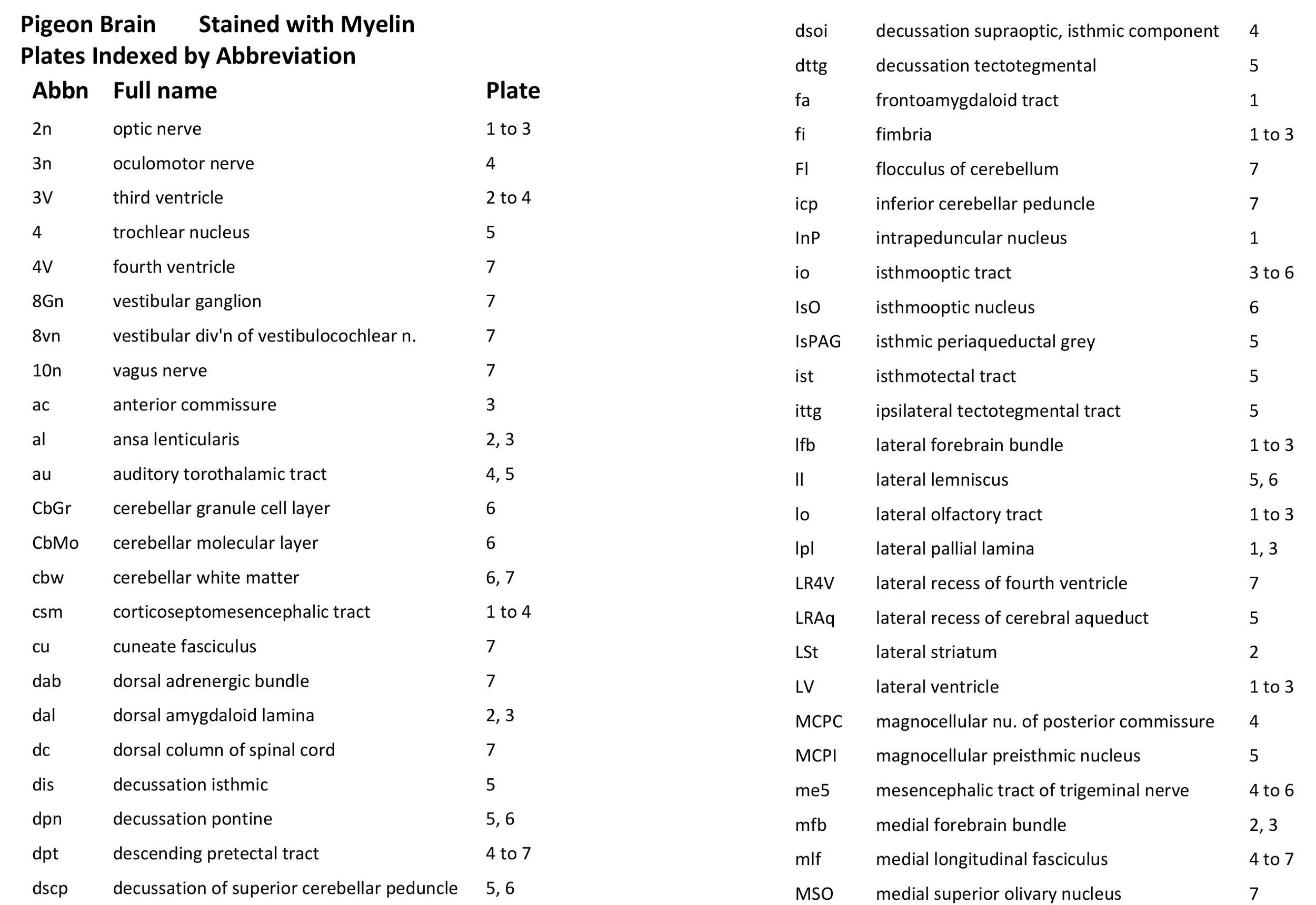
Ansa lenticularis (al) (plates 2, 3) Literally the fibre loop of the lentiform nucleus. The anterior nucleus of the ansa lenticularis has been proposed as the avian homologue of the mammalian subthalamic nucleus (Jiao et al., 2000).
Anterior commissure (ac) (plate 3) This transverse pathway joins the pallia (cortex) of the two cerebral hemispheres and allows interhemispheric transfer of information.
Corticoseptomesencephalic tract (csm) (plates 1 to 4) This prominent bundle connects the medial pallium (cortex) and septal nuclei with the midbrain (mesencephalon). The pathway is also known as the septopalliomesencephlic tract.
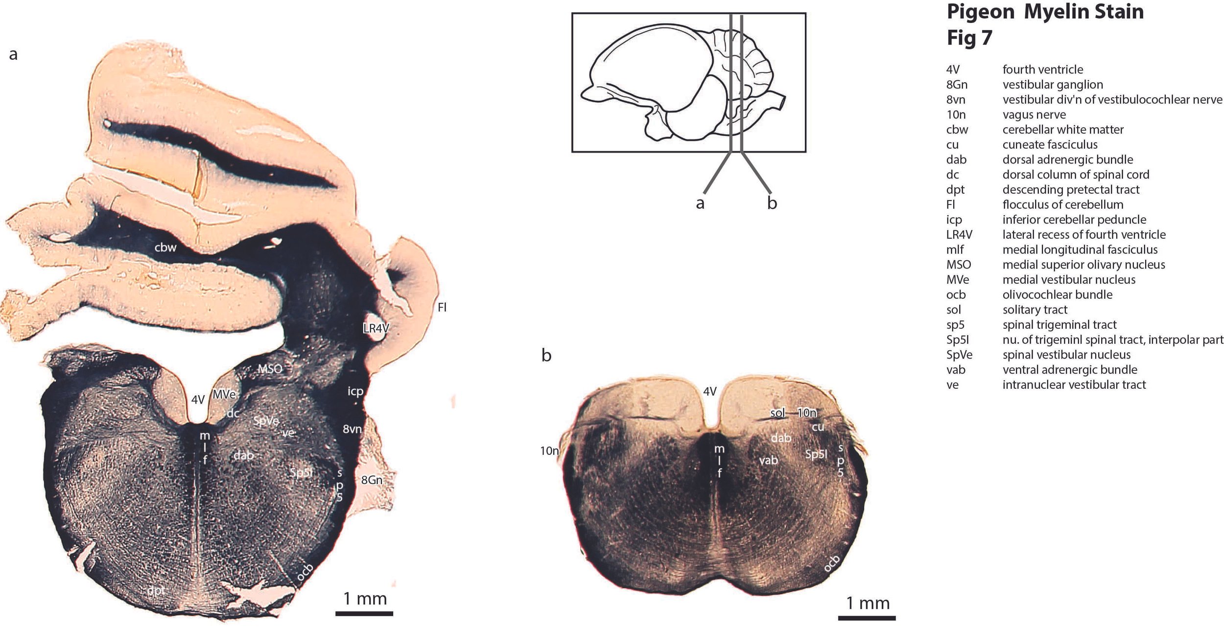
Descending pretectal tract (dpt) (plates 4 to 7) arises in the anterior pretectal nucleus and descends in the ventral tegmentum to the level of the retrotrapezoid nucleus. During its course it passes through the pararubral, pedunculotegmental, descending pretectal tract nucleus, medial pontine, and trapezoid body nuclei.
Dorsal and ventral adrenergic bundles (dab, vab) (plate 7) These longitudinal catecholaminergic pathways are located adjacent to the intermediate reticular formation (IRt), which plays a role in autonomic control of respiratory, cardiovascular and gastrointestinal function.
Frontoamygdaloid tract (fa) (plate 1) arises in the nidopallium and runs rostrocaudally to the amygdala.
Inferior cerebellar peduncle (icp) (plate 7) A fibre bundle mainly carrying proprioceptive information from the spinal cord and vestibular apparatus into the cerebellum. Its terminal part also incorporates the olivocerebellar bundle from the inferior olivary nuclear complex to the cerebellum. The latter is probably critical in training the cerebellum in new motor routines.
Isthmooptic tract (io) (plates 3 to 6) A retinopetal (retina-seeking) pathway which may play a role in focussing visual attention, particularly for the detection of aerial predators (Wilson and Lindstrom, 2011).
Isthmotectal tract (ist) (plate 5) This tract may play a role in reciprocal isthmotectal feedback (i.e. paired tectoisthmic and isthmotectal pathways) and focusses sensory attention on moving objects (Marín et al., 2012).
Lateral forebrain bundle (lfb) (plates 1 to 3) This longitudinally running pathway arises in the subpallium in the vicinity of the basal nucleus of Meynert and passes caudal through the prethalamus in the vicinity of the reticular nuclei, before ending in the dorsal thalamus near the dorsal motor and dorsal somatosensory nuclei.
Medial forebrain bundle (mfb) (plates 2, 3) Also well-known in mammalian brains, this longitudinally running pathway arises in the rostral forebrain between the pallidoseptal transition zone and the nucleus of the horizontal limb of the diagonal band of Broca. It runs caudally through the preoptic area and hypothalamus to the lateral hypothalamic area.
Medial longitudinal fasciculus (mlf) (plates 4 to 7) This prominent tract in both avian and mammalian brains plays a critical role in co-ordination of eye movement. It runs rostrocaudally from the nucleus of Darkschewitsch, to interconnect the oculomotor, trochlear and abducens nuclei. It also receives input from the vestibuloocular pathway and medial vestibular nucleus for vestibular control of eye movement.
Nigrostriatal tract (ns) (plates 2, 3) A dopaminergic pathway from the substantia nigra pars compacta to the neostriatum, seen also in mammals. It facilitates movement via the direct pathway of the palliostriatopallidal circuit and inhibits the indirect pathway.
Oculomotor nerve (3n) (plate 4) The major cranial nerve for control of extraocular muscles and eye movement.
Optic nerve (2n) (plates 1 to 3) A sensory nerve that carries visual information from the eye (in axons of retinal ganglion cells) towards visual centres of the brain (particularly the superficial tectum – see optic tract).
Optic tract (opt) (plates 2 to 6) Once retinal ganglion cell axons have passed the optic chiasm (och) they enter the optic tract and terminate principally in the superficial layer of the tectum.
Posterior commissure (pc) (plate 4) This is a major commissure located at the caudal end of prosomere 1 (pretectum). One of its major roles is to allow transmission of information between pretectal reflex centres on each side of the brain, ensuring that pupillary reflexes are symmetrical for both eyes.
Quintofrontal tract (qf) (plates 2, 3) Also known as the quintofrontal tract of Wallenberg, this is a direct monosynaptic ascending projection from the principal trigeminal sensory nucleus to the nucleus basalis of the telencephalon. It stands in contrast to other vertebrates where trigeminal somatosensory information is processed in the dorsal thalamus (e.g. mammalian VPM nucleus) before ascending to the telencephalon. The pathway has been implicated in the sensorimotor and motivational control of feeding (Zeigler, 1973).
Tectal grey commissure (tgc) (plate 4) A robust commissure interconnecting the visual centres of the tectum on each side.
Tectothalamic tract (tth) (plate 4, 5) An ascending pathway from auditory midbrain to the auditory dorsal thalamus. This pathway includes an inhibitory (GABAergic) projection from the torus semicircularis to the auditory thalamus (Ito and Atoji, 2016).
Tectotegmental tract (ttg) (plate 4) This includes tectopontine, tectoreticular and crossed tectobulbar descending pathways (Reiner and Karten, 1982).
References
Puelles L, Martinez-de-la-Torre M, Paxinos G, Watson C, Martinez S (2007) The Chick Brain in Stereotaxic Coordinates. An Atlas featuring Neuromeric Subdivisions and Mammalian Homologies. Academic Press.
Hutchins B, Weber JT (1983) A rapid myelin stain for frozen sections: modification of the Heidenhain procedure. J Neurosci Methods 7: 289-294.
Ito T, Atoji Y (2016) Tectothalamic inhibitory projection neurons in the avian torus semicircularis. J Comp Neurol 524 10.1002/cne.23979
Jiao Y, Medina L, Veenman CL, Toledo C, Puelles L, Reiner A (2000) Identification of the anterior nucleus of the ansa lenticularis in birds as the homolog of the mammalian subthalamic nucleus. J Neurosci 20: 6998-7010.
Marín GJ, Durán E, Morales C, Gonzalez-Cabrera C, Sentis E, Mpodozis J, Letelier JC (2012) Attentional capture? Synchronised feedback signals from the isthmi boost retinal signals to higher visual areas. J Neurosci 32: 1110-1122.
Reiner A, Karten HJ (1982) Laminar distribution of the cells of origin of the descending tectofugal pathways in the pigeon (Columba livia). J Comp Neurol 204: 165-187.
Wilson M, Lindstrom SH (2011) What the bird’s brain tells the bird’s eye: the function of descending input to the avian retina. Vis. Neurosci 28: 337-350.
Zeigler HP (1973) Trigeminal deafferentation and feeding in the pigeon: sensorimotor and motivational deficits. Science 18: 155-159