Atlas of the Brain and Head of the Tammar Wallaby (Notamacropus eugenii) at P5 (haematoxylin and eosin)
-
The tammar wallaby (Notamacropus eugenii) is a small macropod originally found in Western and South Australia and on ten or more offshore islands. Wild tammars usually inhabited coastal scrub and dry sclerophyll forest. The species breeds well in captivity, making it an ideal diprotodontid marsupial for developmental studies.
In the wild, pouch-young are usually born in late January (mid-summer) after a gestation of 26.5 days. Females can mate again within a few hours of the initial birth, but the conceptus from the second mating usually remains quiescent during lactation. In the natural environment the pouch-young are suckled for 8 to 9 months and leave the pouch in September or October.
-
The wallabies depicted in these series of colour plates were obtained from a breeding colony at the Australian National University (ANU) in the Australian Capital Territory (ACT). Some monochrome images of these specimens have been shown in a previous publication (Ashwell et al., 2010).
All experimental procedures were approved by the Animal Ethics Experimentation Committee of the ANU, conform to NIH principles of laboratory animal care and were carried out according to the ethical guidelines of the National Health and Medical Research Council (Australia). The age of the pouch young animal was determined either directly by noting the elapsed time from the date of birth, which was designated P0, or from measurements of head-length and reference to a chart of head-lengths of animals of known age. This is accurate to within approximately 2 days.
The pouch-young tammars were anaesthetized by hypothermia and perfused with normal saline followed by Bouin’s fixative. The heads were stored in 70% ethanol prior to embedding in paraffin and sectioning coronally at a thickness of 10 µm. The sections were mounted on subbed glass slides and stained with haematoxylin and eosin before being coverslipped with DePeX.
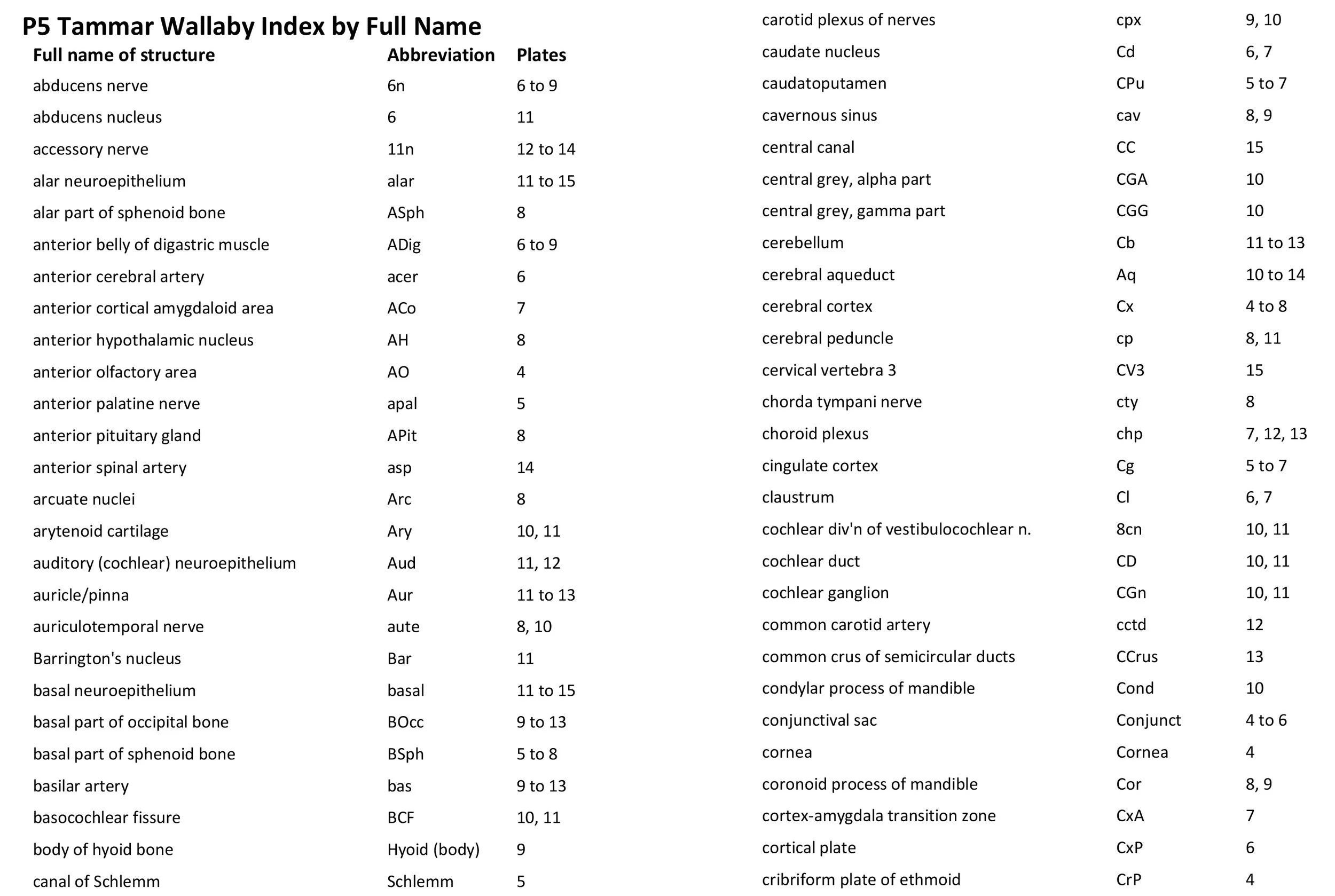
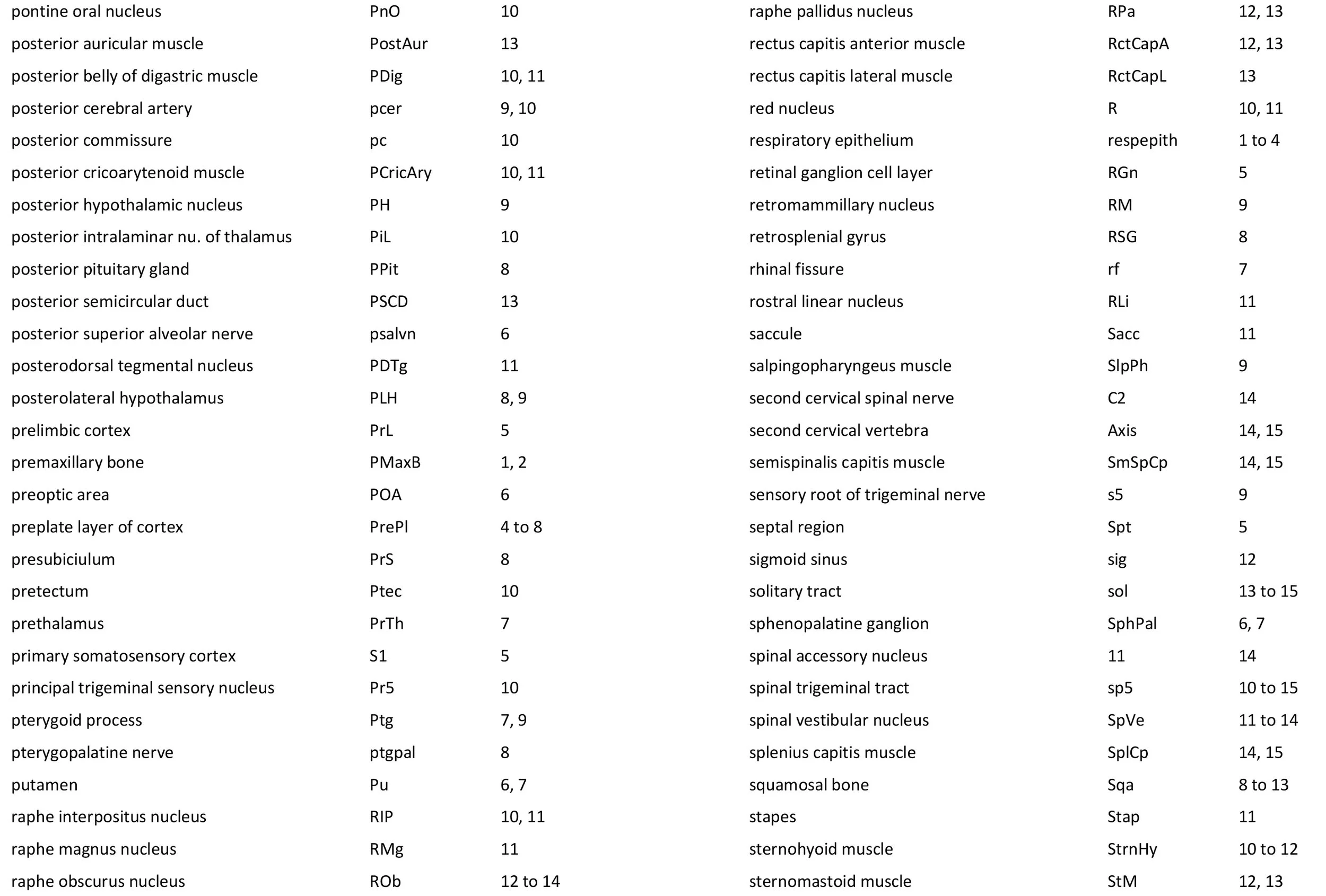
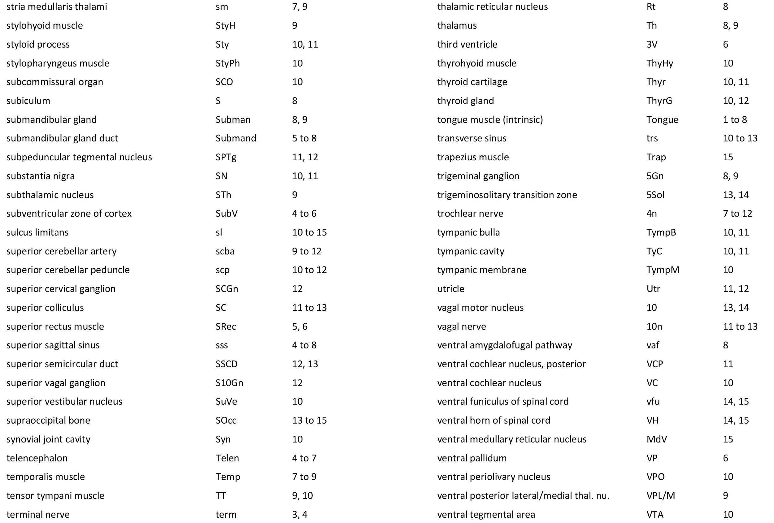
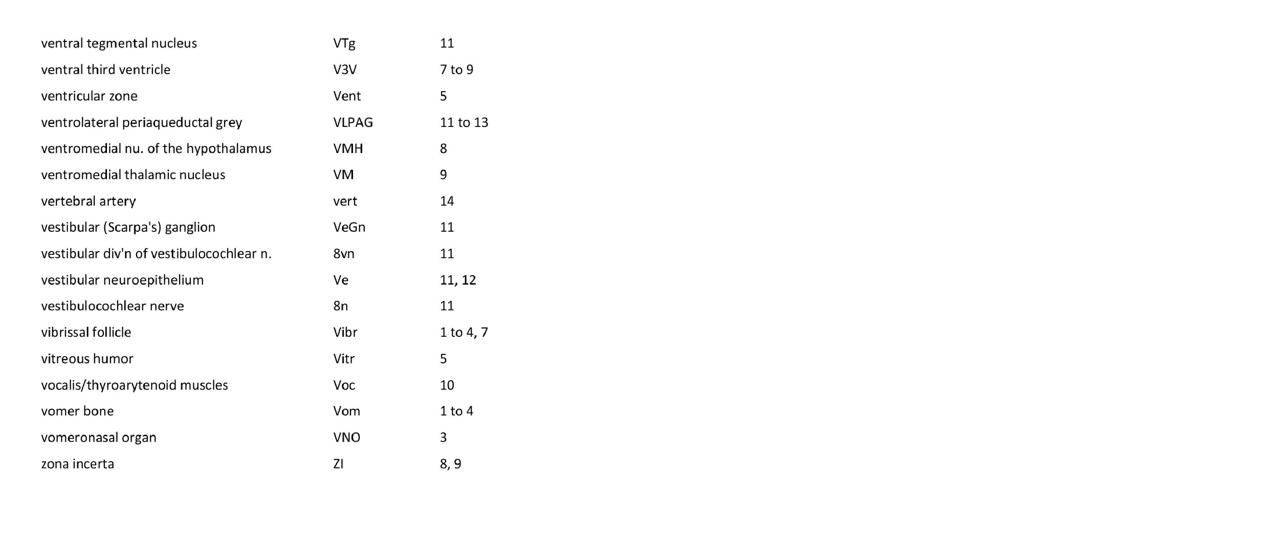
Each section was photographed with the aid of a Zeiss Axiophot photomicrographic system and a Canon EOS 400D body. Images were placed in Adobe Illustrator 2021 and delineated. Developmental regions (i.e. neuroepithelium) destined to give rise to adult structures have been denoted by the adult structure’s name with an asterisk (e.g. Cb* denotes the cerebellar developmental field of the rhombic lip).
The heads are essentially bilaterally symmetrical, so only half the head has been depicted, with a 1 mm scale bar indicating the size of structures in the dehydrated tissue. A small finder cartoon has been provided in each plate to show the rostrocaudal position of the section. The distance from the rostral tip of the snout can be calculated by multiplying the section number by 10 µm.
-
Ashwell KWS, Marotte LR, Mai JK (2010) Atlas of the brain of the developing tammar wallaby (Macropus eugenii). In: The Neurobiology of Australian Marsupials. (K Ashwell, ed). Cambridge: Cambridge University Press.
Plates 1-15 of a tammar wallaby pouch young at P5
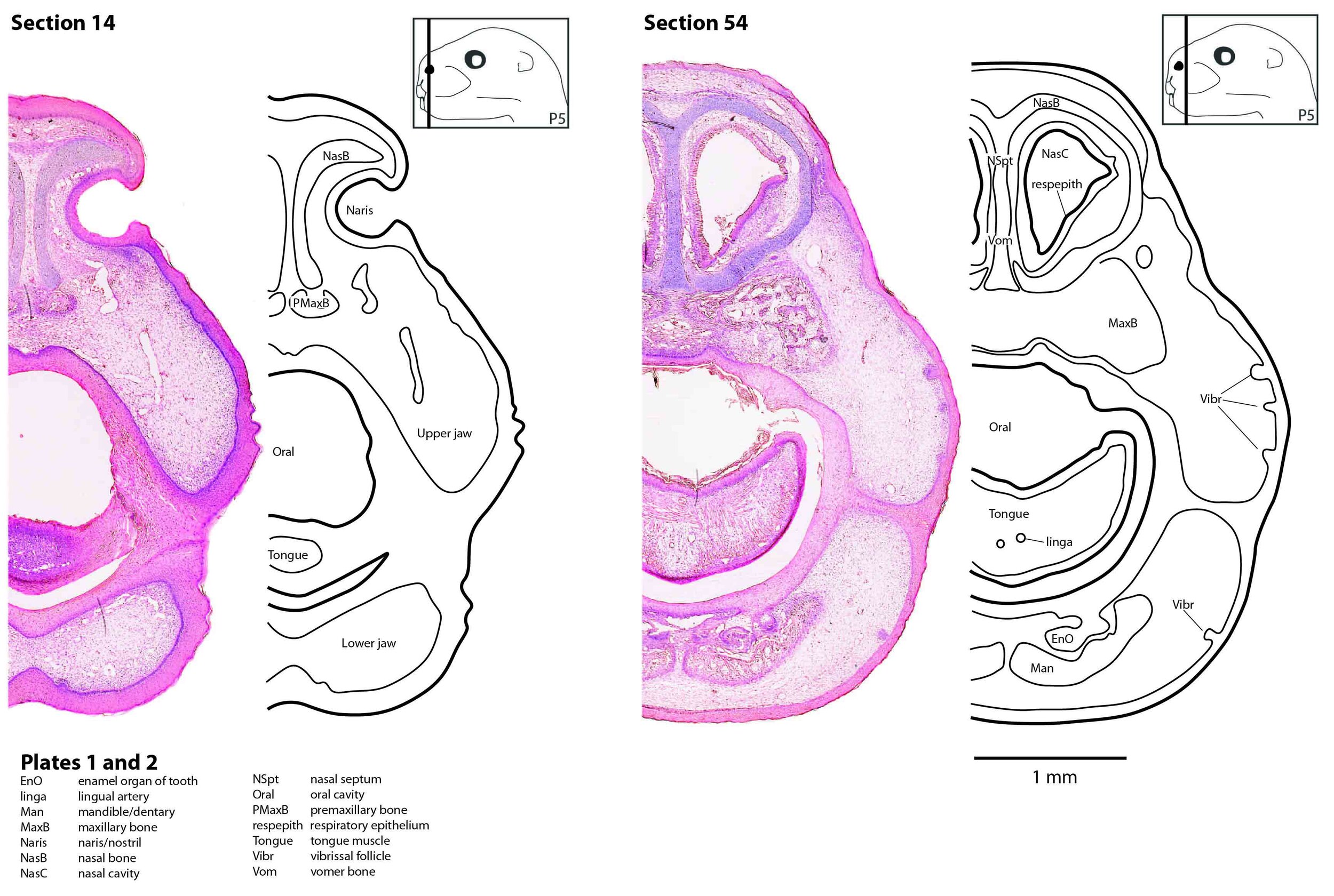
Figure 1 shows sections 14 and 54 through the head of a P5 tammar wallaby pouch young. These sections are 140 and 540 µm from the tip of the snout, respectively.
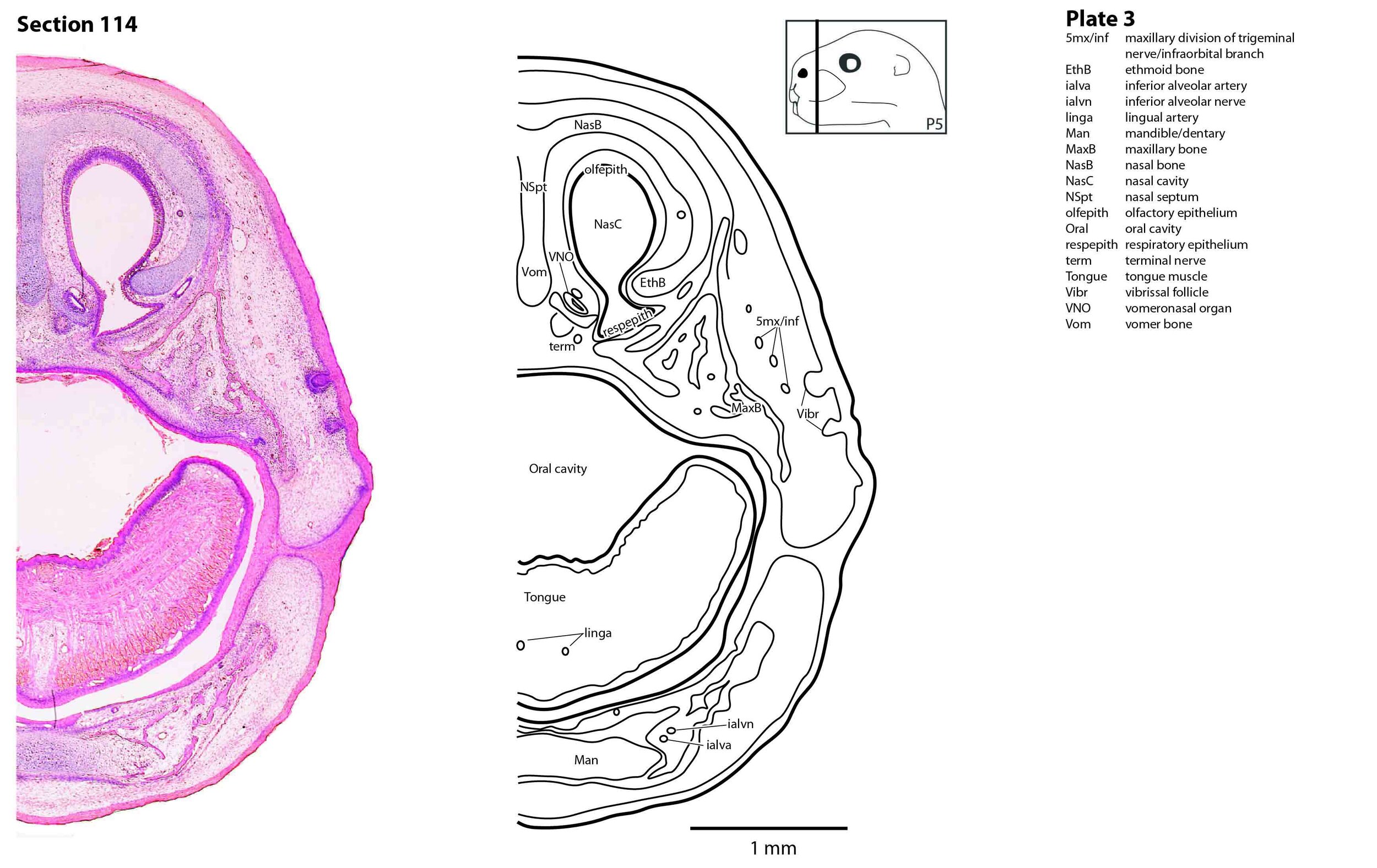
Figure 2 shows section 114 through the head of a P5 tammar wallaby pouch young. This section is 1140 µm from the tip of the snout.
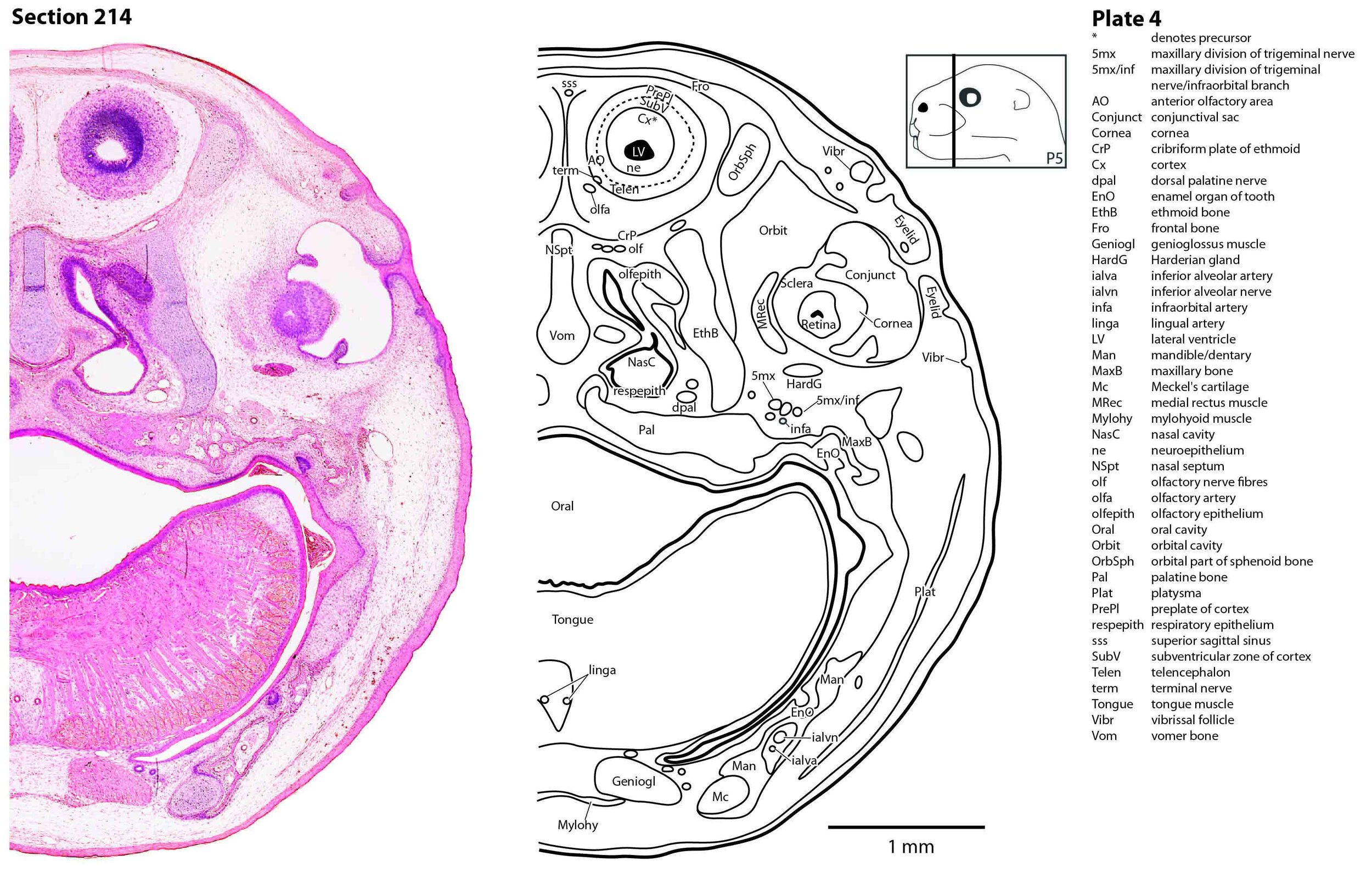
Figure 3 shows section 214 through the head of a P5 tammar wallaby pouch young. This section is 2140 µm from the tip of the snout.

Figure 4 shows section 254 through the head of a P5 tammar wallaby pouch young. This section is 2540 µm from the tip of the snout.
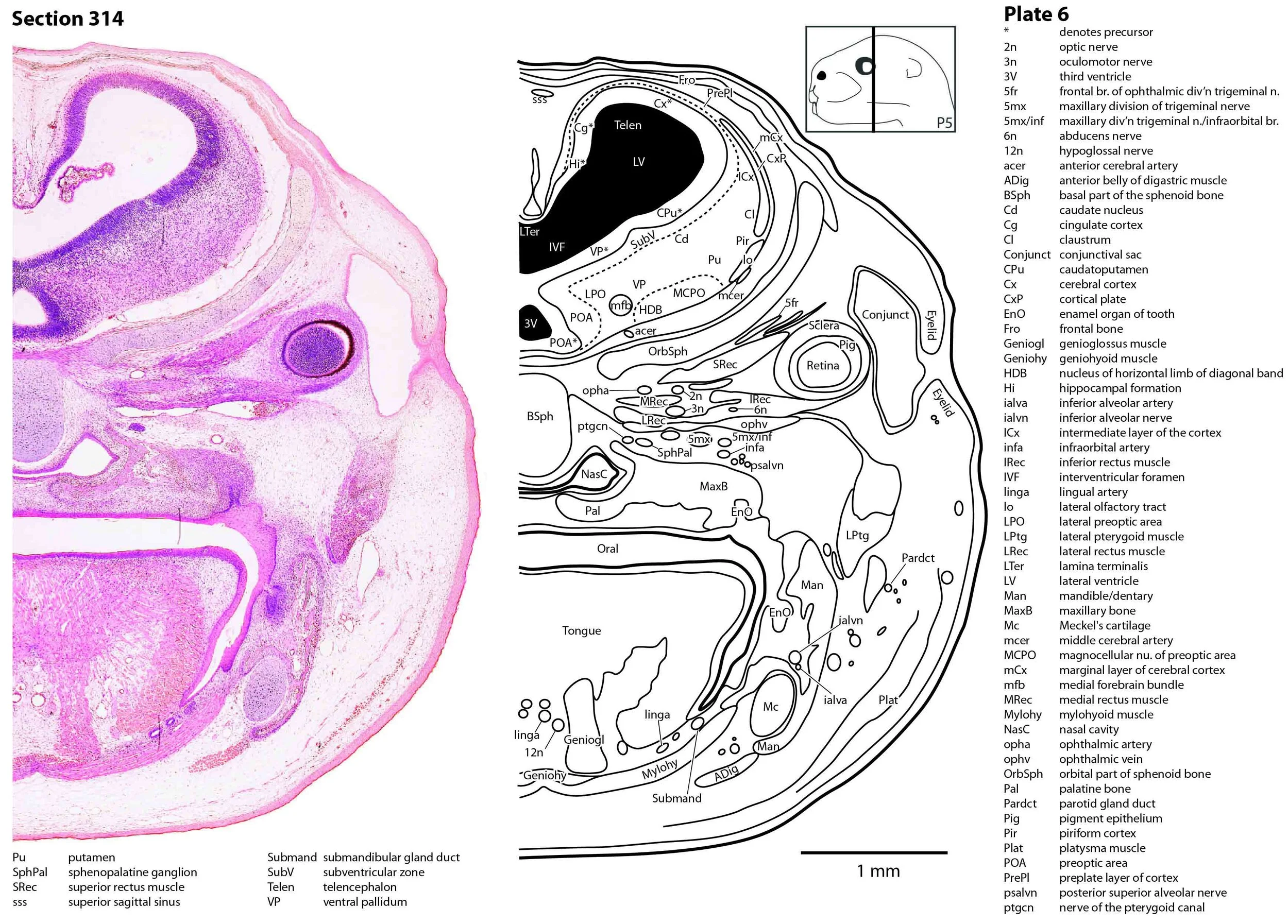
Figure 5 shows section 314 through the head of a P5 tammar wallaby pouch young. This section is 3140 µm from the tip of the snout.

Figure 6 shows section 354 through the head of a P5 tammar wallaby pouch young. This section is 3540 µm from the tip of the snout.

Figure 7 shows section 414 through the head of a P5 tammar wallaby pouch young. This section is 4140 µm from the tip of the snout.
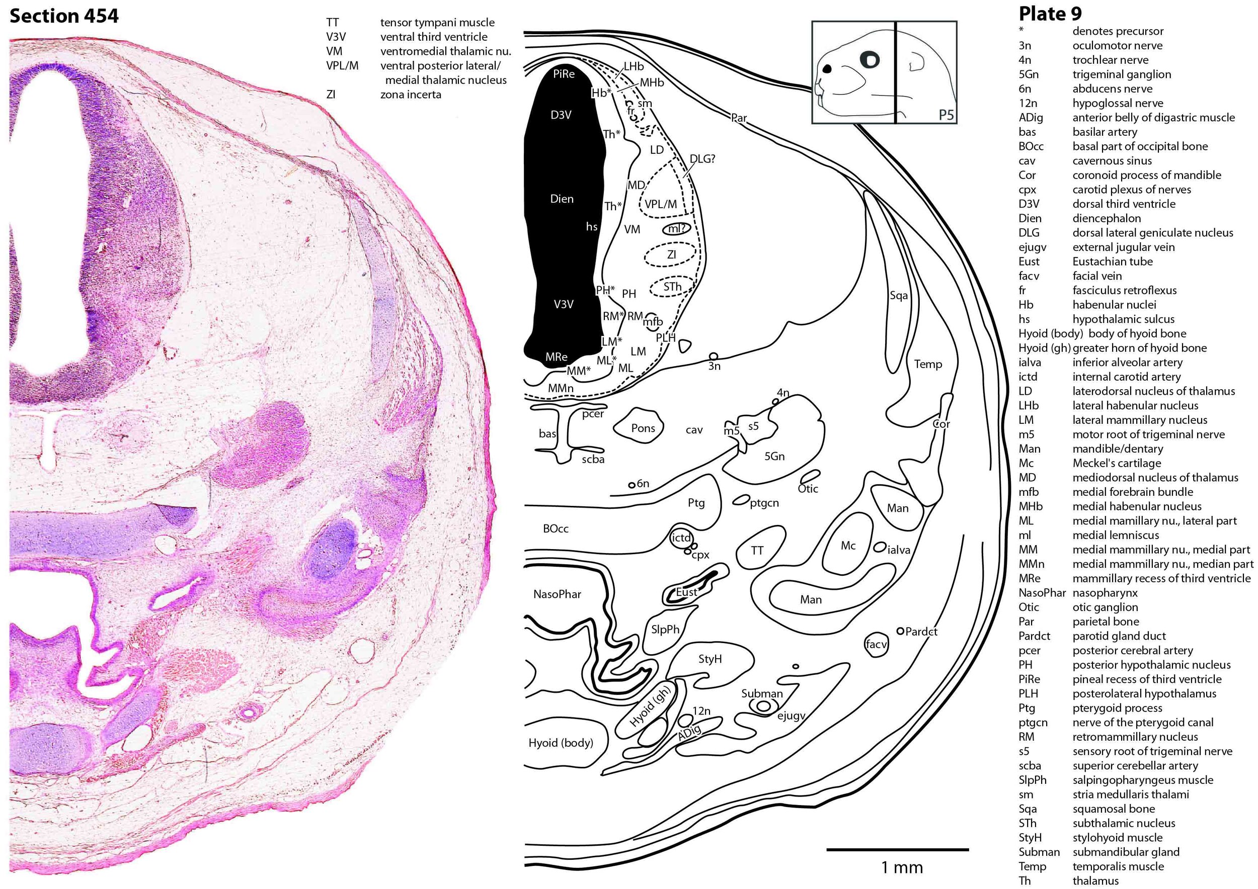
Figure 8 shows section 454 through the head of a P5 tammar wallaby pouch young. This section is 4540 µm from the tip of the snout.

Figure 9 shows section 514 through the head of a P5 tammar wallaby pouch young. This section is 5140 µm from the tip of the snout.
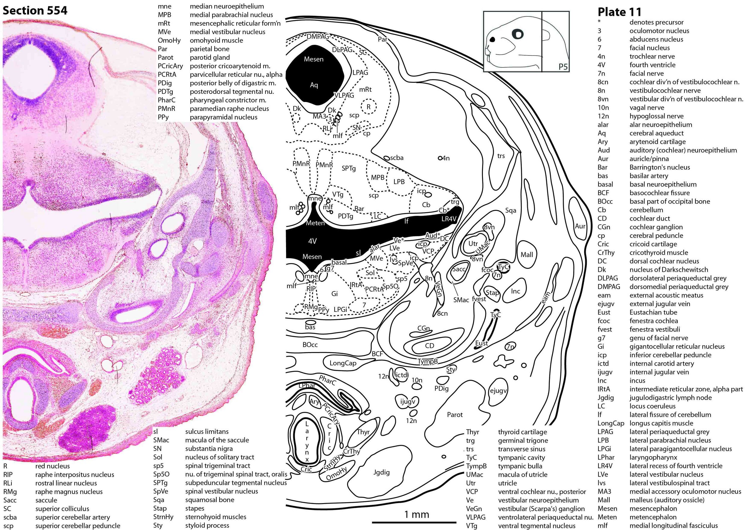
Figure 10 shows section 554 through the head of a P5 tammar wallaby pouch young. This section is 5540 µm from the tip of the snout.

Figure 11 shows section 614 through the head of a P5 tammar wallaby pouch young. This section is 6140 µm from the tip of the snout.
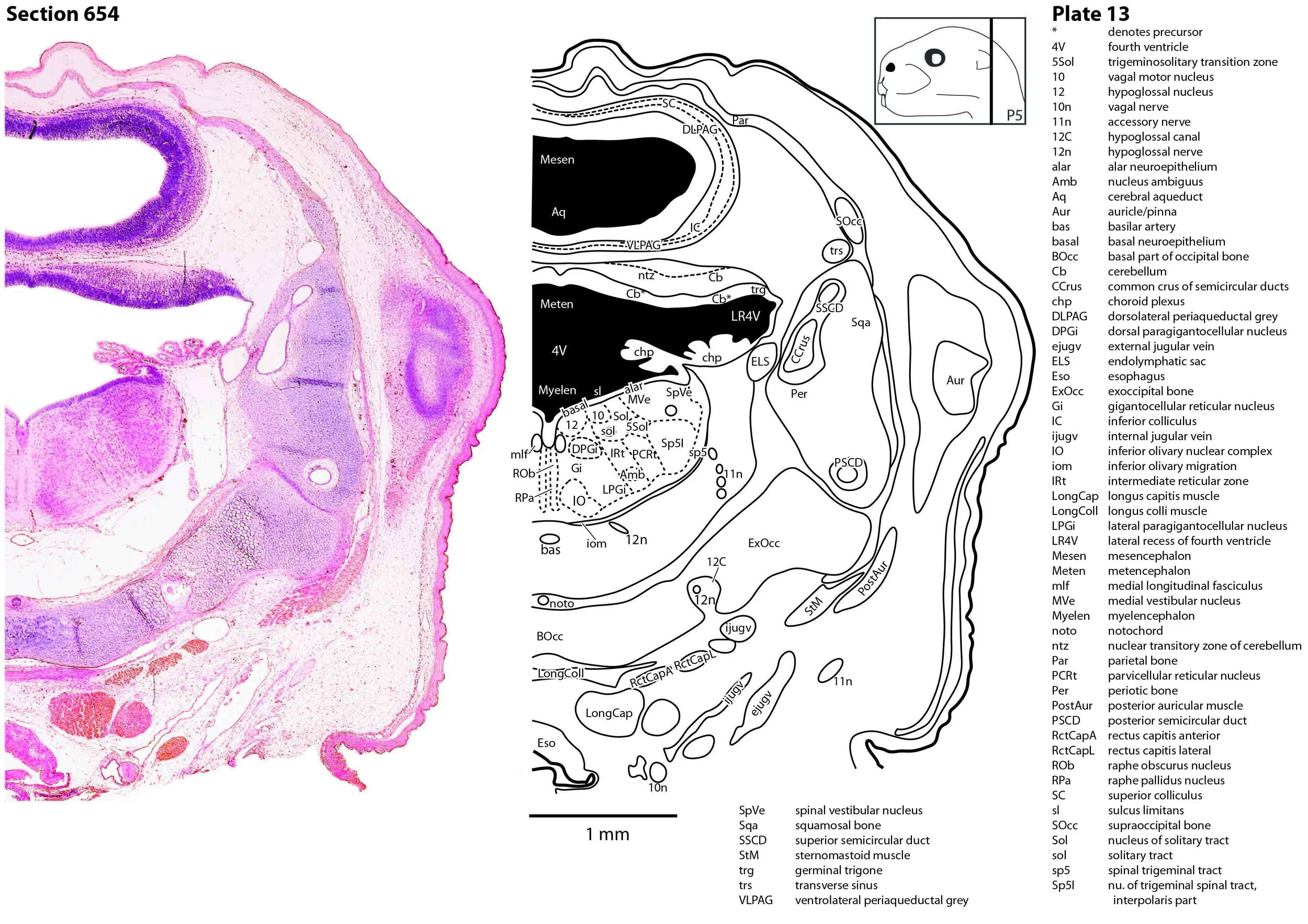
Figure 12 shows section 654 through the head of a P5 tammar wallaby pouch young. This section is 6540 µm from the tip of the snout.

Figure 13 shows section 714 through the head of a P5 tammar wallaby pouch young. This section is 7140 µm from the tip of the snout.
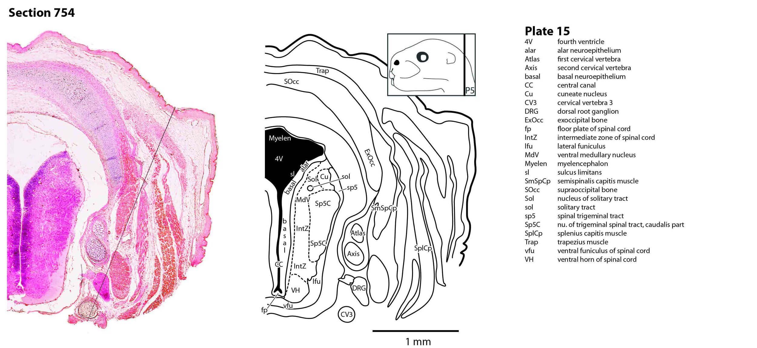
Figure 14 shows section 754 through the head of a P5 tammar wallaby pouch young. This section is 7540 µm from the tip of the snout.