Atlas of the Head of a Newborn Brushtail Possum (MS289)
Introduction
The common brushtail possum (Trichosurus vulpecula) is a nocturnal, semi-arboreal, phalangerid, diprotodontid marsupial native to the eastern and south-eastern parts of Australia (including Tasmania), pockets of Western Australia, some offshore islands (e.g. Kangaroo and Barrow), the Kimberley, Arnhem Land, and also invasive in New Zealand. Brushtail possums have also adapted well to Australian urban environments, raiding rubbish and compost bins. Adult possums can weigh up to 4.5 kg and have a bushy tail that is prehensile and naked on its ventral side. Their usual diet is omnivorous, including eucalyptus leaves, flowers, shoots, fruits and seeds. On occasion they may also eat eggs, insects and small vertebrates to supplement protein intake.
Newborn brushtail possums weigh 2 g and are 15 mm in length. Gestation is 18 days, followed by 4 to 5 months of postnatal life in the forward-facing pouch, and a further 4 to 5 months riding on the mother’s back.
Methods
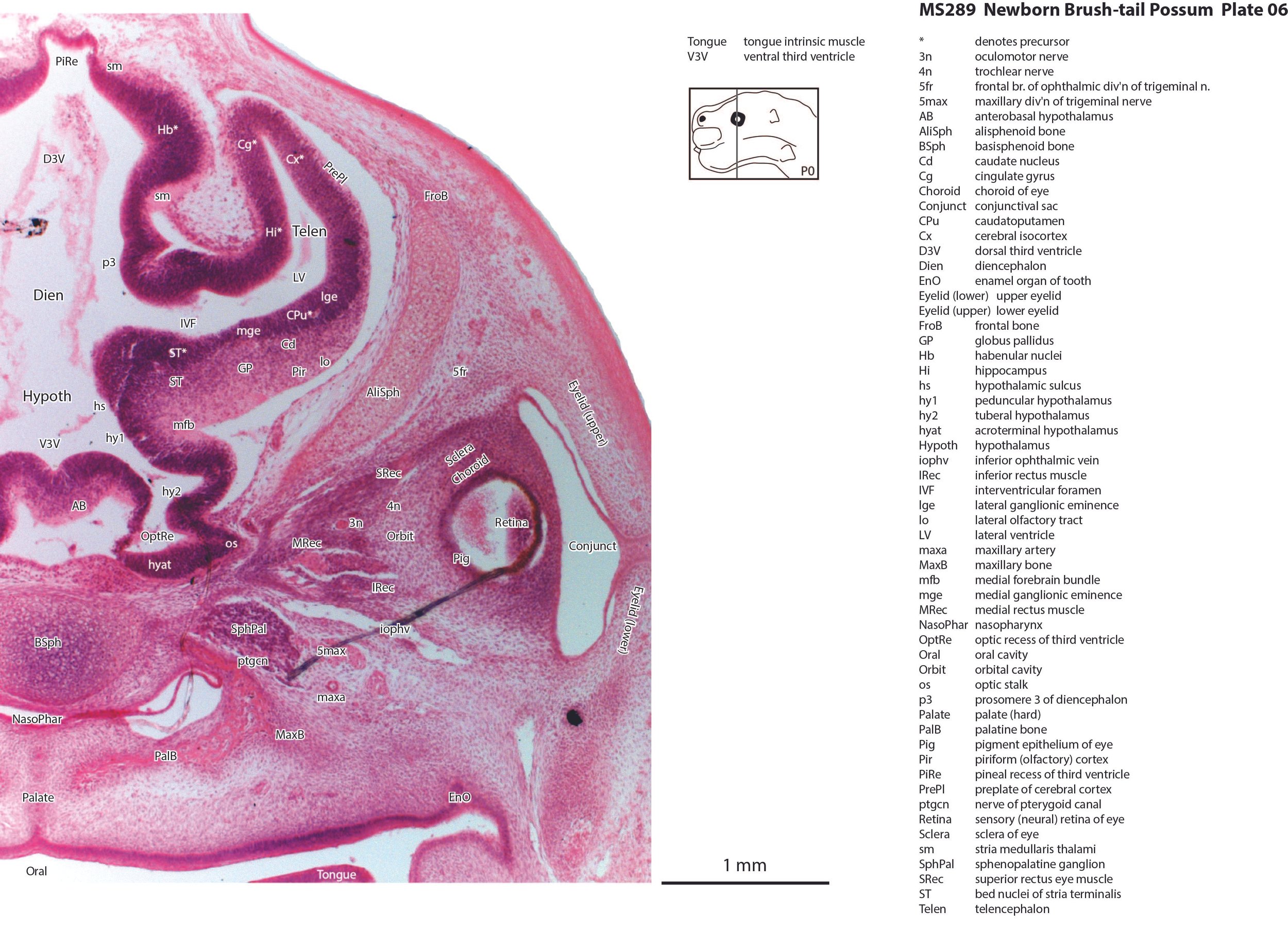
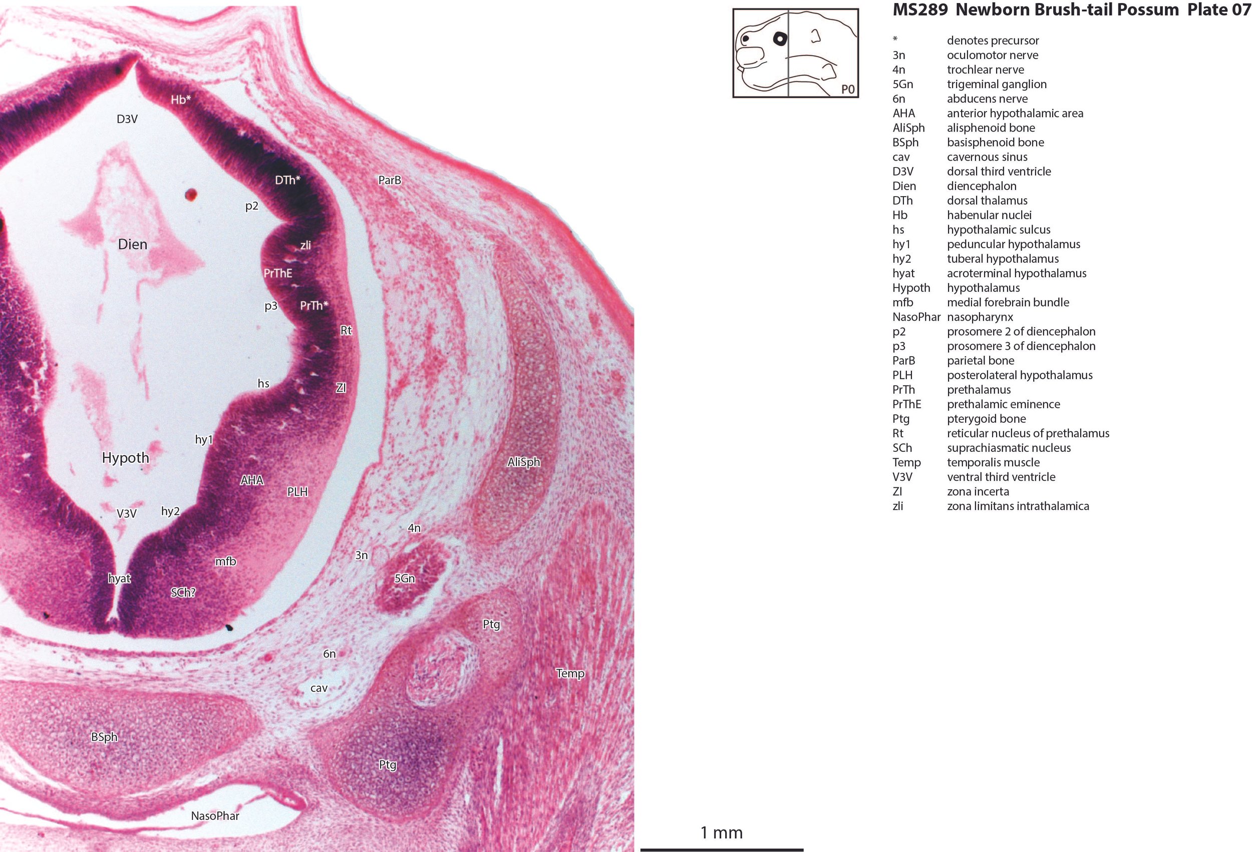
The specimen illustrated here is MS289 (Trichosurus vulpecula, female newborn pouch young, greatest length of 15 mm, head length of 6.5 mm) of the Hill collection stored at the Museum für Naturkunde in Berlin.
The specimen was collected in 1897, embedded in paraffin wax and sectioned transversely at 15 µm thickness, before being stained with hematoxylin and eosin.
Sections at approximately 150 µm intervals were photographed with the aid of a Zeiss Axioplan2 fitted with an AxioCam MRc5 camera. Sections at 300 µm intervals have been depicted in the plates and the section numbers are consecutive from the snout tip. All images were calibrated by photographing a scale bar at the same magnification. Images were placed in Adobe Illustrator 2023 and delineated. Developmental regions (i.e. neuroepithelium) destined to give rise to adult structures have been denoted by the adult structure’s name with an asterisk (e.g. Cx* denotes the developmental field of the cerebral isocortex).
An image of the profile of a newborn possum head was not available, so a line diagram of the head of a newborn tammar wallaby has been used for the finder illustration indicating section position.
Notes on the specimen
General observations
There is a notable thickening of the epidermis (plates 1, 2) across the snout, maxilla and rostral jaw, representing the oral shield (Schneider and Gurovich, 2017).
Central nervous system
The olfactory bulb (plate 4) is still rudimentary, with a very thick generative (mitotic) neuroepithelium and very few postmitotic neurons released. The telencephalon has a thin pallium (Cx, Cg and Hi), with a thin external preplate (plates 4 to 6) over the dorsal and medial pallium. The lateral cortex (e.g. Pir in plates 4 to 6) is better developed, but also thin and immature. The hippocampus (plates 5, 6) is also rudimentary, i.e it has a thick proliferative zone, surmounted by a thin preplate zone, which is mostly fibrous with very few neurons. The subpallial telencephalon has the usual lateral ganglionic eminence (lge in plates 5, 6) and medial ganglionic eminence (mge in plate 5, 6). The striatal subventricular zone has not yet developed and although there are some early-generated striatal and pallidal neurons, it is not possible to distinguish internal structure among those neuronal populations. Only the notional positions of striatal and pallidal components have been labelled.
The hypothalamus is divided into peduncular (hy1), tuberal (hy2), and acroterminal (hyat) proliferative components (all in plates 6, 7). The rostral parts of the hypothalamus (plate 6) are very rudimentary and have essentially no postmitotic neurons external to the proliferative zones. In more caudal sections (plate 7) there are more postmitotic neurons in putative nuclei, including suprachiasmatic and posterolateral hypothalamic nuclei. There is also a diffuse medial forebrain bundle (mfb in plates 6 to 8), running longitudinally through the nascent hypothalamic nuclei.
The diencephalon has the usual prosomeric structure. Prosomeres 1 (pretectum), 2 (dorsal thalamus), and 3 (prethalamus) can be identified as proliferative zones, but postmitotic populations are very small, particularly in the dorsal thalamus.
The mesencephalon has a rudimentary superior colliculus (SC in plates 10 to 13) and inferior colliculus (IC in plates 12, 13), where most of the neuraxis wall is made up of the proliferative neuroepithelium, and very few postmitotic neurons. The mesencephalic tegmentum is also undeveloped. Only the notional position of major nuclei can be marked, because differentiation of nuclei and fibre tracts is poor.
The cerebellum (plates 10 to 13) is a lid extending over the fourth ventricle and rimmed by the rostral rhombic lip. No cortical differentiation and only limited deep nuclear differentiation (putative lateral deep cerebellar nucleus) is evident.
The brainstem of this newborn possum is at a similar level of differentiation to the newborn tammar wallaby. The trigeminal sensory nuclear complex (Pr5, Sp5O, Sp5I; plates 9 to 14; Sp5C not shown) is easily distinguished and the medial magnocellular reticular formation (Gi, GiV, LPGi; plates 10 to 14) is broader that the parvicellular reticular formation (PCRt; plates 11 to 14). This would be consistent with a major role for trigeminal somatosensation in analysing the environment and feeding information to the brainstem reticulospinal pathways for co-ordination of forelimb movement. A putative lateral vestibulospinal tract (lvs in plates 12 to 14) is identified and may mediate control of spinal cord motoneurons in response to vestibular input.
Peripheral nervous system
Most cranial nerves can be identified. Cranial nerve 1 is multiple fibres (olf in plate 3) running from the olfactory epithelium to the nascent olfactory bulb. Cranial nerve 2 (optic) is not developed, but an optic stalk (os in plate 6) extends from the hypothalamus to the eye cup. The eye muscle nerves 3n, 4n and 6n can be traced from the brainstem to the orbit (3n in plates 5 to 10, 4n in plates 5 to 13, 6n in plates 7, 8). The trigeminal nerve (5n in plate 9) runs from the trigeminal ganglion (5Gn in plates 7 to 9) to the brainstem. Distal from the ganglion, the maxillary division and its distal branch 5max/inf (plates 1 to 6) is the most obvious of the three trigeminal divisions. The facial nerve can be traced from the facial motor nucleus (7 in plate 11) through the (internal) genu of the facial nerve (g7 in plate 10) and externally as 7n (plate 12). Components of the vestibulocochlear nerve pathway include the cochlear ganglion (CGn in plate 11), vestibular ganglion (VeGn in plates 11 and 12), and centrally as the vestibulocochlear nerve (8n in plate 11) and its vestibular division (8vn in plate 11). The emergent glossopharyngeal/vagal/accessory nerve rootlets are visible as a group, but only the vagus nerve (10n in plate 13) and superior vagal ganglion (S10Gn in plate 13) can be picked out clearly. The hypoglossal nerve (12n) emerges from the ventral brainstem (plates 12 to 14) and exits the hypoglossal canal (plate 13).
Sense organs
The olfactory epithelium of the nasal cavity (olfepith in plates 2 to 4) is thicker than respiratory epithelium (respepith in plates 2 to 4). The eye structure is sufficiently developed that cornea (plate 5), lens (plate 5), sensory retina (plates 5, 6), and pigment epithelium (Pig in plates 5, 6) can be distinguished, but the retina is still in an extremely primitive state with few or no postmitotic neurons. The inner ear has progressed to a stage where cochlear duct (plate 11), utricle (plates 11, 12), saccule (plates 11, 12), semicircular ducts (plates 11 to 13) and endolymphatic duct and sac (plates 13, 14) can be identified, but sensory regions (maculae in plates 11, 12) are still extremely immature. Mystacial vibrissae (Vibr in plates 2, 3) are in the process of differentiation, but the follicles consist only of an epidermal peg surmounted by dermal condensation. Some simple early innervation is likely, but not visible with this stain (see tammar GAP-43 atlas).
Musculoskeletal system
Cartilaginous models for the premaxilla, nasal, ethmoid, lacrimal, maxillary, presphenoid, basal sphenoid, basal occipital, squamosal, periotic, ex- and supraoccipital bones are present.
Acknowledgements
I would like to thank Dr Peter Giere of the MfN, Berlin Germany, for access to the collection and for all his kind help during the work.
References
Schneider NY, Gurovich Y (2017) Morphology and evolution of the oral shield in marsupial neonates including the newborn monito del monte (Dromiciops gliroides, Marsupialia Microbiotheria) pouch young. J Anat 231, 59-83.