Cytoarchitectural Atlas of the Brain of a Pigeon (Columba livia domestica)
Introduction
Feral or urban pigeons (Columba livia domestica or Columba livia forma urbana) are descendants of domestic pigeons that have returned to the wild and become pests in modern cities. The bird is derived from the wild rock dove of Europe, North Africa, Middle East and India.
Methods
This specimen was painlessly euthanised with pentobarbitone sodium and the brain removed and immersed in formal saline for 48 hours. The brain was subsequently immersed in phosphate-buffered 30% sucrose for 24 hours and sectioned frontally on a freezing microtome at a thickness of 50 µm. Selected sections were either mounted onto gelatinized glass slides and stained for Cresyl violet (current series) or stained free-floating for myelin with a modified Heidenhain technique (see myelin series).
The sections were photographed with a Canon 4000D and an EFS 60 mm FL macrolens. Where necessary, images were photomontaged with Adobe Photoshop 2023 and labelled with Adobe Illustrator 2023.
Nomenclature and delineation were based on a detailed atlas of the chick brain (Puelles et al., 2007), because the brains of the chicken and pigeon are very similar. Readers are referred to that work for detailed explanations of neuromeric subdivision in the mature avian brain.
Notes on the specimen
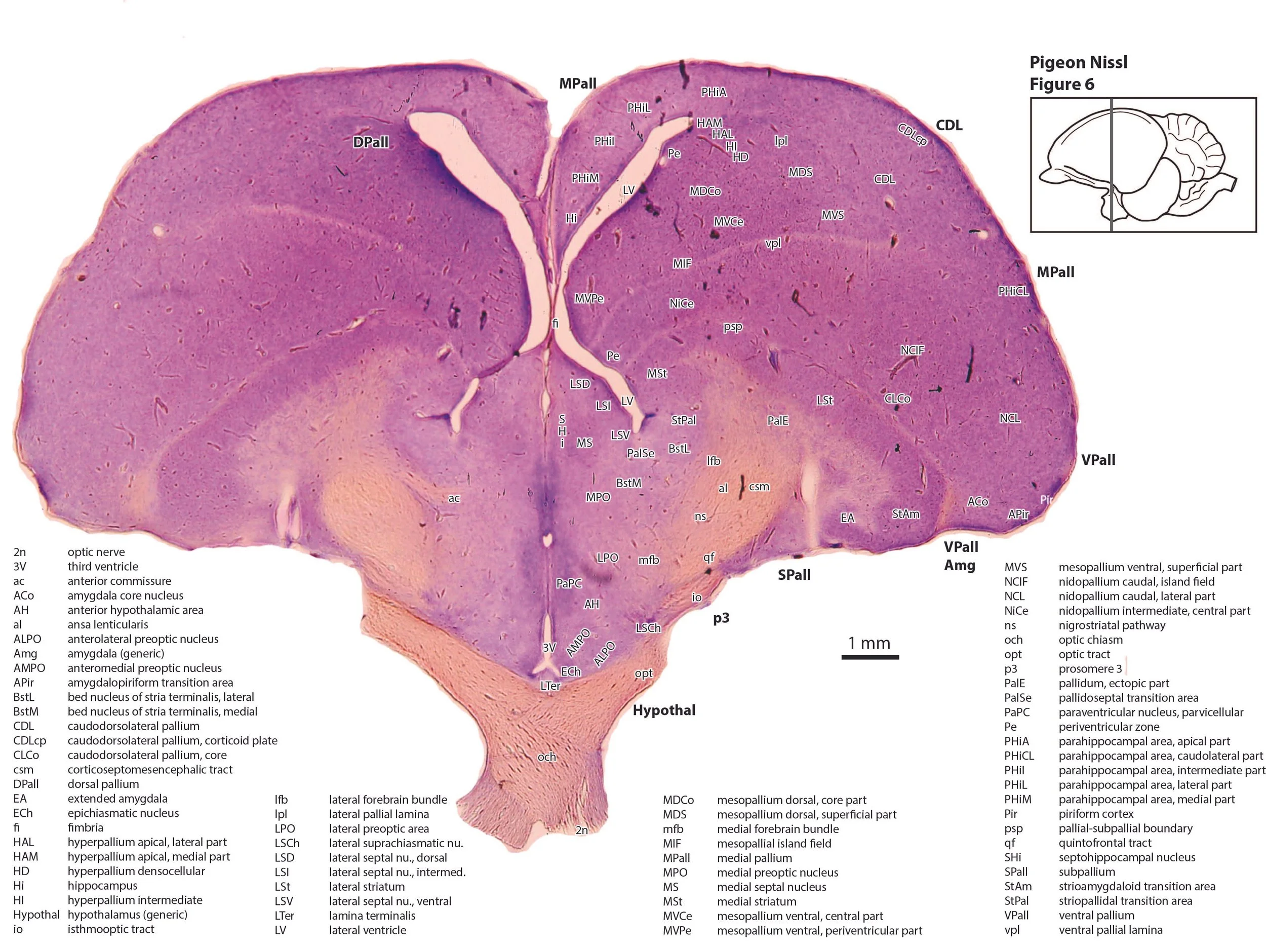
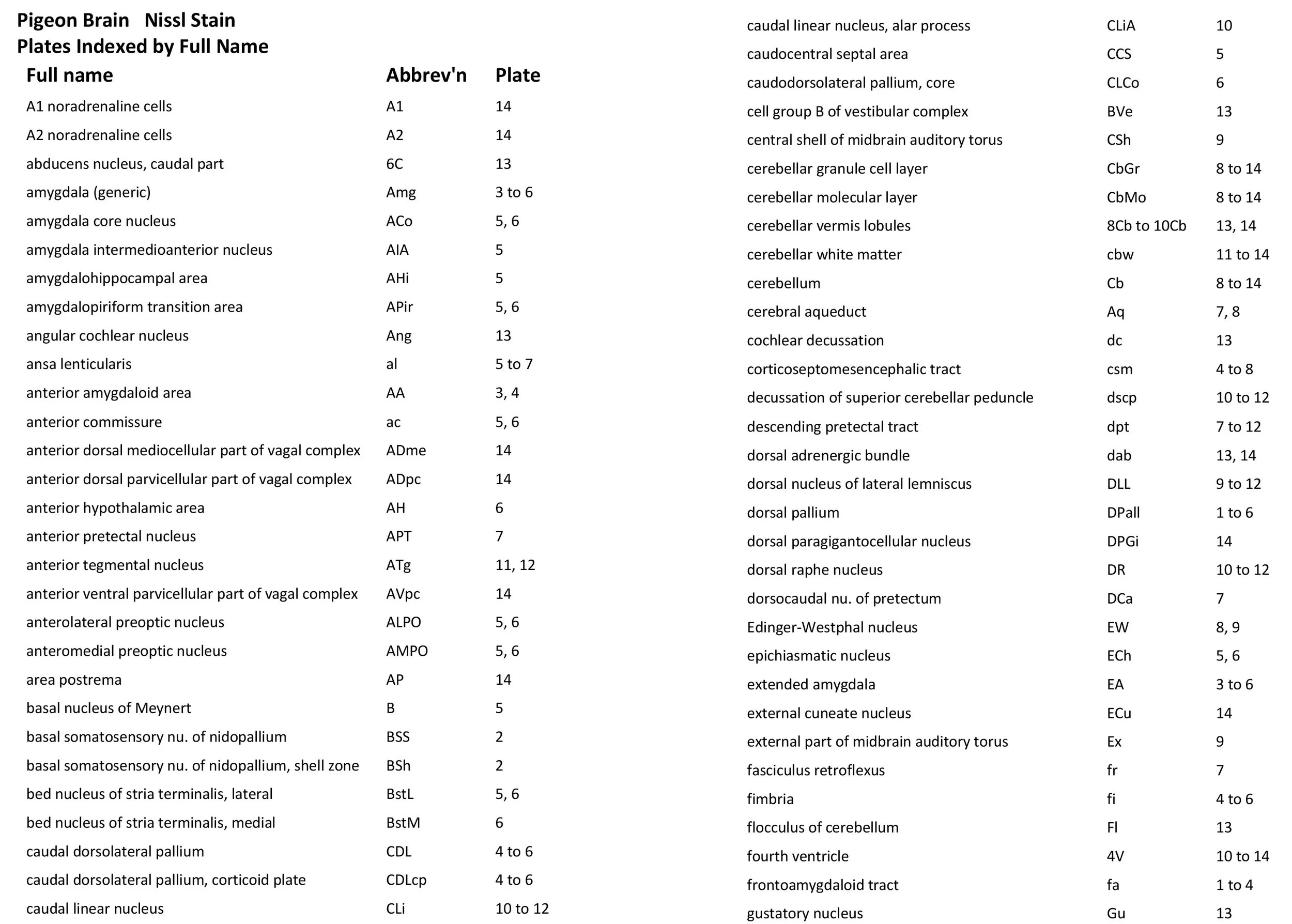
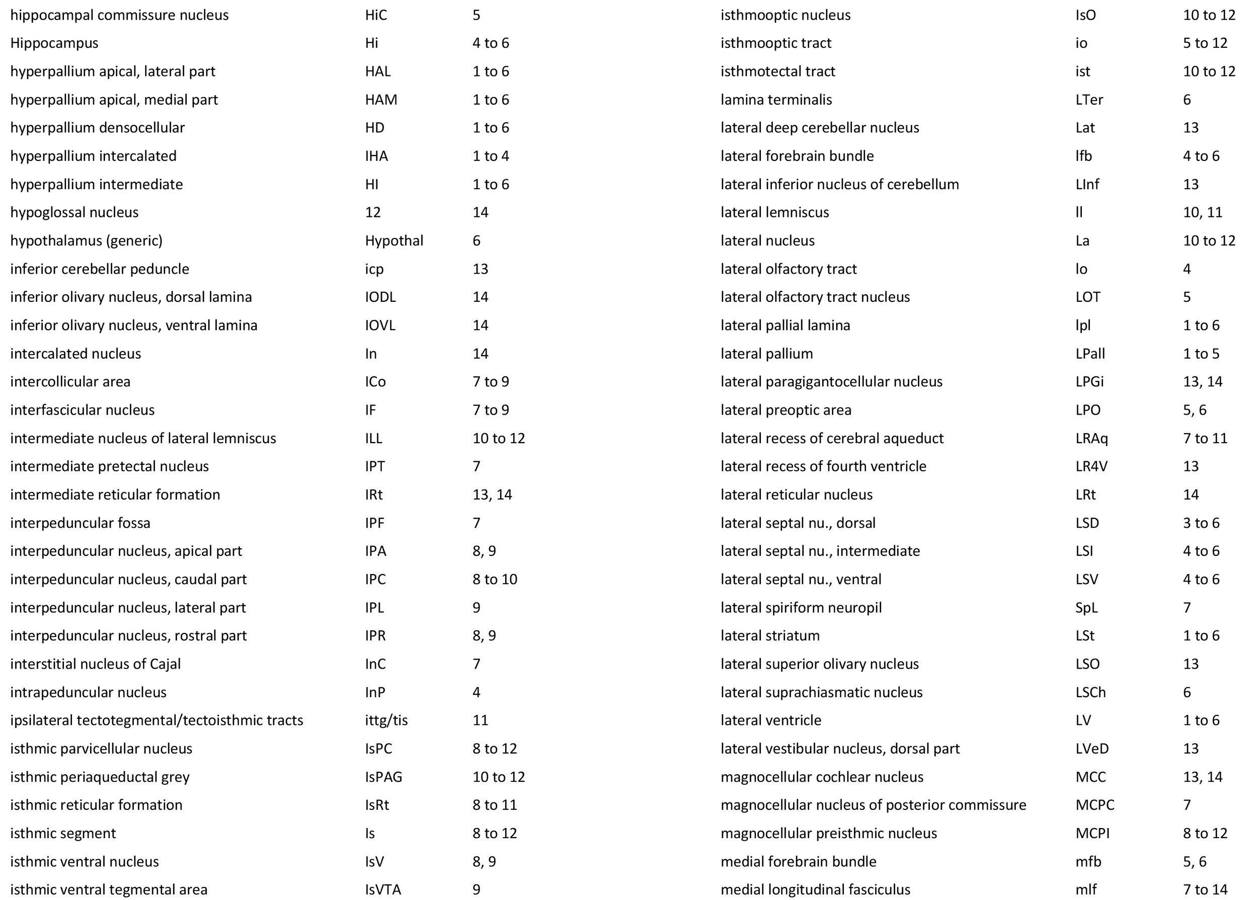
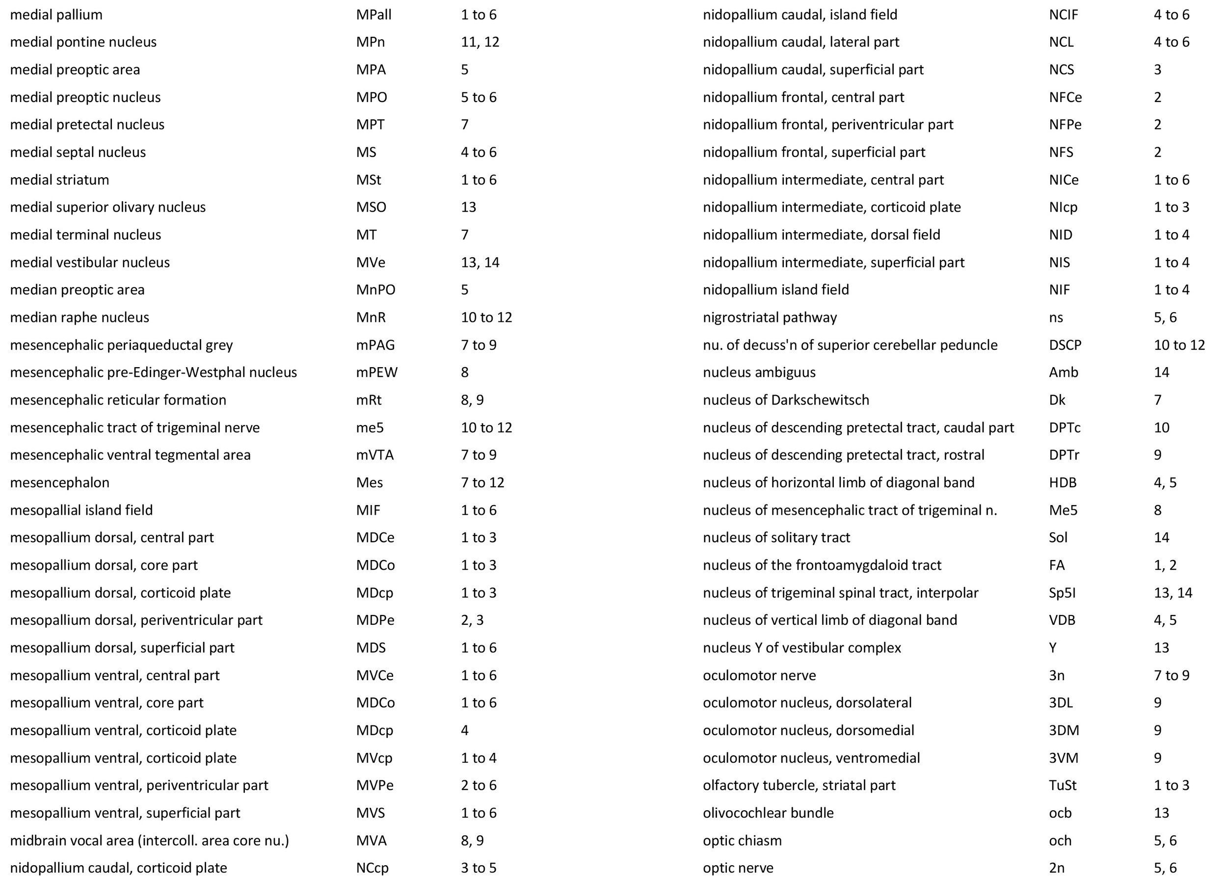
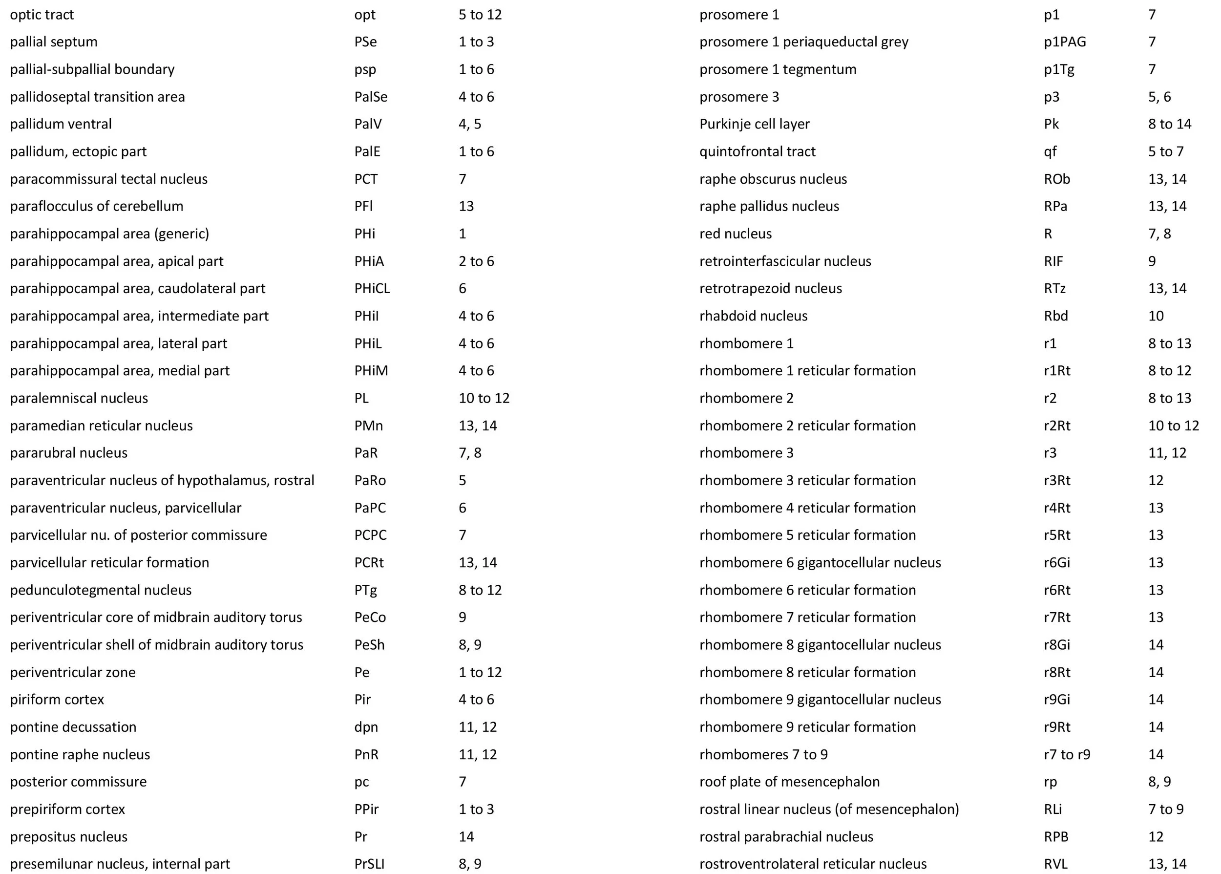
The sections depicted in plate 1 to 14 are not in a regular sequence, so please refer to the finder image in each for the section level. Figure 1 depicts a line diagram of the lateral view of a typical pigeon brain showing the main external features.
A key difference between avian and mammalian brains is that the avian pallium is formed into rather homogenous, densely populated neuronal groups (hyperpallium, mesopallium, nidopallium), rather than the laminated and often folded pallium (cortex) of mammals. The external surface of the avian forebrain is divided into mesopallium (including parahippocampal, entorhinal and hippocampal regions), dorsal pallium, lateral pallium, caudodorsolateral pallium, ventral pallium and subpallium. Within the pallial interior are hyperpallial, mesopallial, and nidopallial zones, arranged from dorsal to ventral and separated by white matter laminae. The hyperpallium is separated from the mesopallium by the lateral pallial lamina (lpl in plates 1 to 6), the mesopallium is separated from the nidopallium by the ventral pallial lamina (vpl in plates 1 to 6), and the nidopallium is separated from the subpallium by the pallial-subpallial boundary (psp in plates 1 to 6).
The nidopallium contains the functionally important basal somatosensory nucleus and its surrounding shell (BSS and BSh, respectively, in plate 2), as well as the prominent visual nidopallial core and shell (VisCo, VisSh in plates 1 to 4).
The midbrain features a large visual tectum (Tect in plates 7 to 12) and a deeper auditory torus semicircularis (ToS in plates 8, 9). The isthmo-optic nucleus (IsO in plates 10 to 12) gives rise to a retinopetal pathway called the isthmo-optic tract (io in plates 5 to 12), which may play a role in focussing visual attention for aerial predators (Wilson and Lindstrom, 2011).
The bulk of the avian cerebellum is made up of the vermis (Cb in plates 8 to 14), with small laterally projecting flocculus and paraflocculus (Fl and PFl, respectively, in plate 13).
The vagal motor complex of the medulla for visceral control is extensive, including ventral lateral (VLa), anteroventral (AVpc) and anterodorsal medio- and parvicellular (ADme, ADpc) components (see plate 14).
References
Puelles L, Martinez-de-la-Torre M, Paxinos G, Watson C, Martinez S (2007) The Chick Brain in Stereotaxic Coordinates. An Atlas featuring Neuromeric Subdivisions and Mammalian Homologies. Academic Press.
Wilson M, Lindstrom SH (2011) What the bird’s brain tells the bird’s eye: the function of descending input to the avian retina. Vis. Neurosci 28: 337-350.