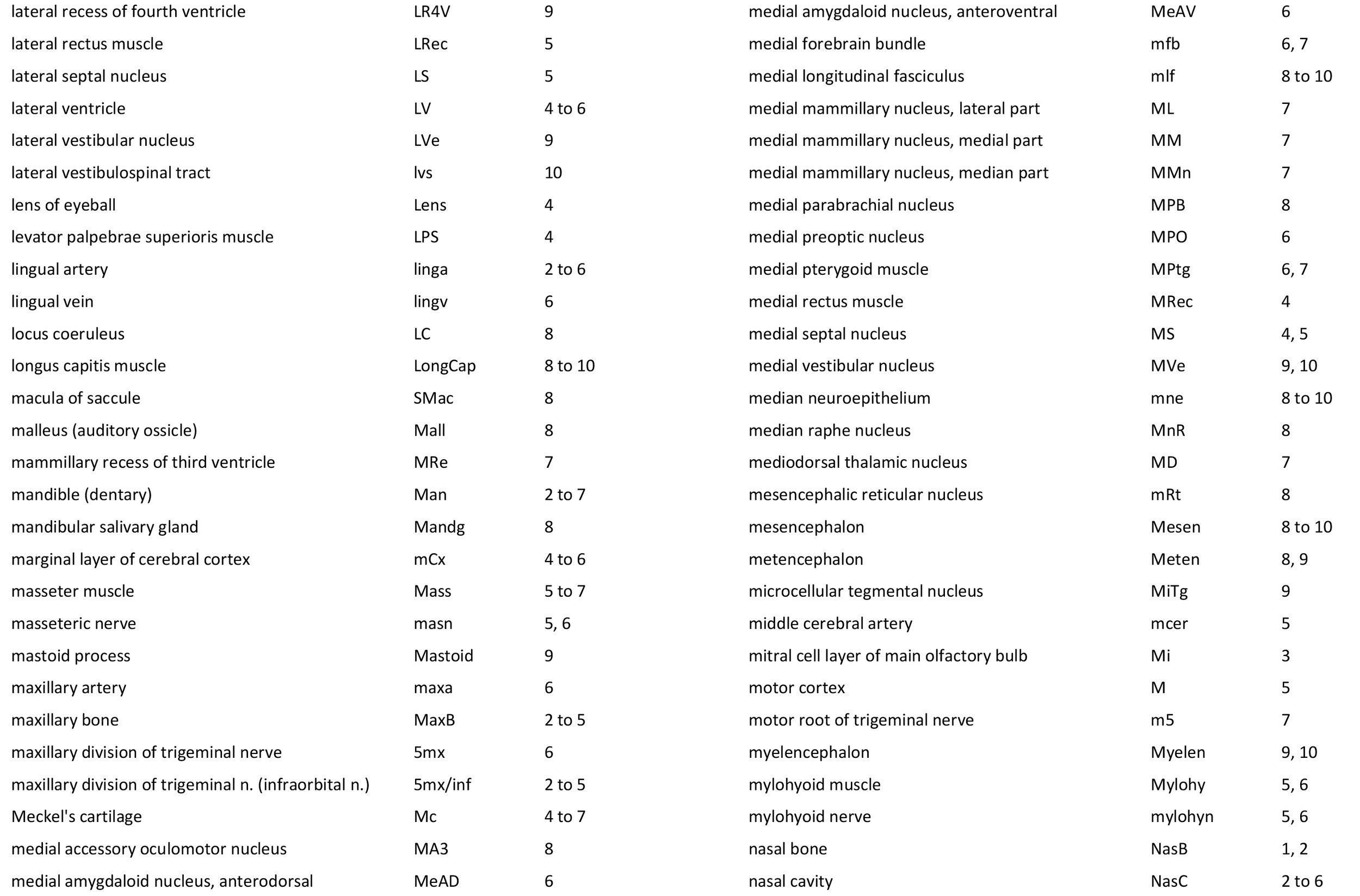
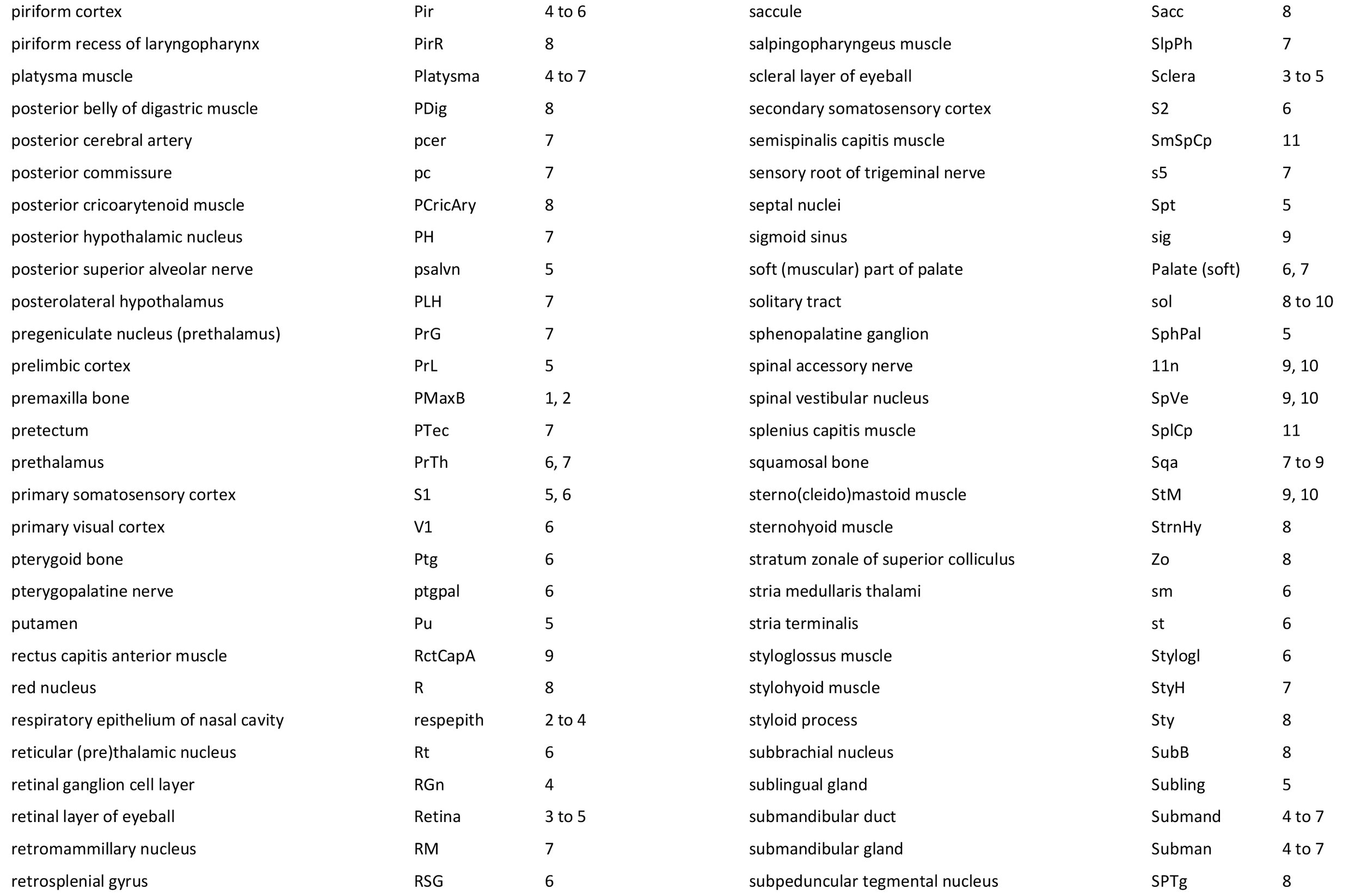
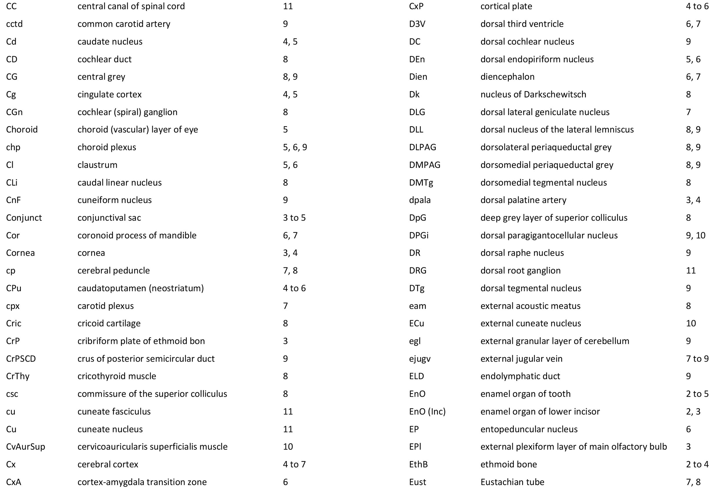
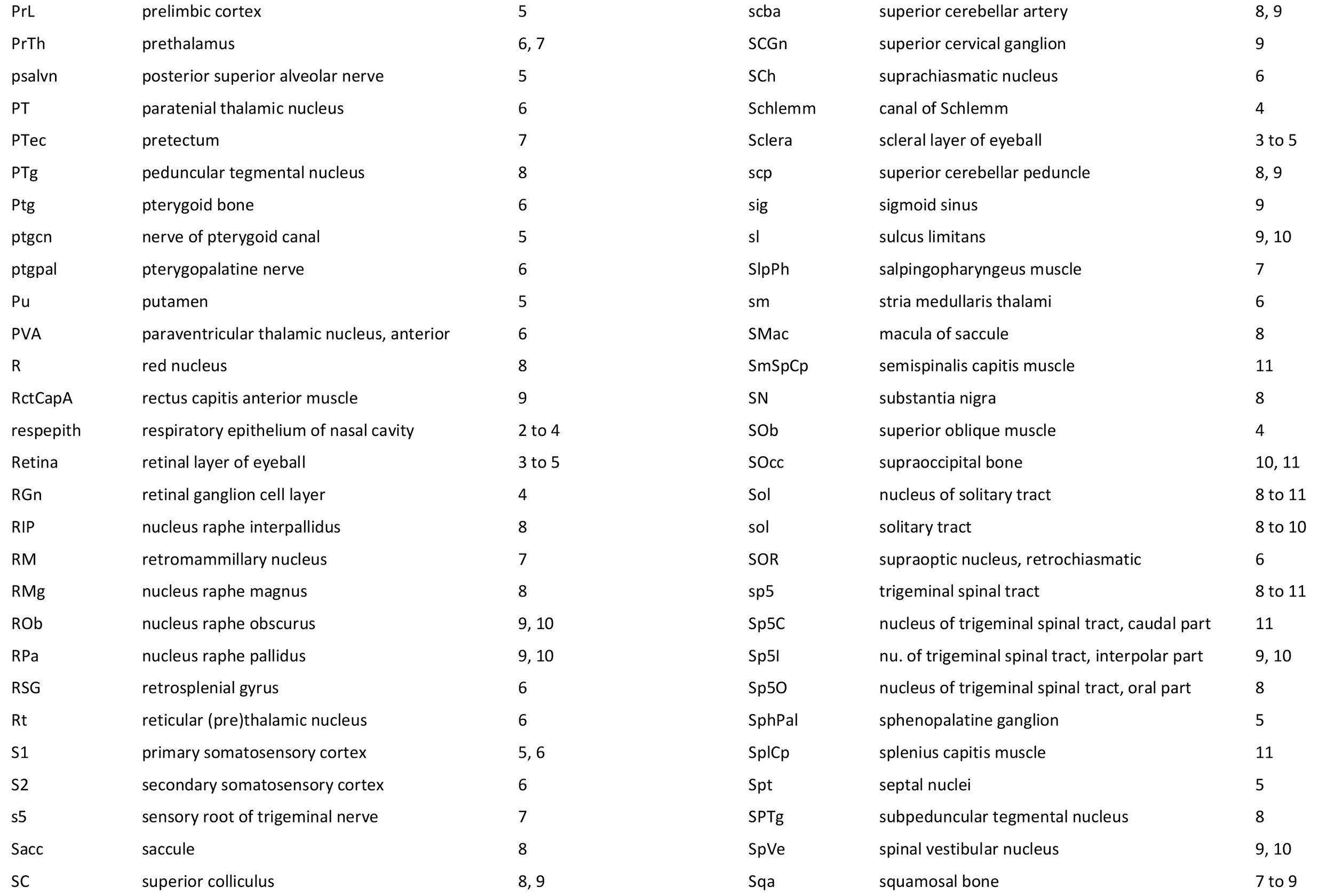
Atlas of the Brain and Head of the Tammar Wallaby (Notamacropus eugenii) at P12 (haematoxylin and eosin)
The tammar wallaby (Notamacropus eugenii) is a small macropod originally found in Western and South Australia and on ten or more offshore islands. Please see P5 tammar atlas for further details.
Methods
The wallaby depicted in the series of colour plates was obtained from a breeding colony at the Australian National University (ANU) in the Australian Capital Territory (ACT). Some monochrome images of this specimen have been shown in a previous publication (Ashwell et al., 2010). The animal’s head was approximately 11.5 mm in rostrocaudal extent. The head was embedded in paraffin and sectioned at a thickness of 10 µm in the laboratory of Prof Dr Jürgen K. Mai at Heinrich Heine Universität, Düsseldorf.
Please see P5 atlas for further details.
Each section was photographed with the aid of a Zeiss Axiophot photomicrographic system and a Canon EOS 400D body. Images were placed in Adobe Illustrator 2021 and delineated. Developmental regions (i.e. neuroepithelium) destined to give rise to adult structures have been denoted by the adult structure’s name with an asterisk (e.g. Cb* denotes the cerebellar developmental field of the rhombic lip).
The head is essentially bilaterally symmetrical, so only half the head has been depicted, with a 1 mm scale bar indicating the size of structures in the dehydrated tissue. A small finder cartoon has been provided in each plate to show the rostrocaudal position of the section. The distance from the rostral tip of the snout to the section depicted can be calculated by multiplying the section number by 10 µm.
References
Ashwell KWS, Marotte LR, Mai JK (2010) Atlas of the brain of the developing tammar wallaby (Macropus eugenii). In: The Neurobiology of Australian Marsupials. (K Ashwell, ed). Cambridge: Cambridge University Press.
Figure 1 shows section 34 through the head of a P12 tammar wallaby pouch young. This section is 340 µm from the tip of the snout.
Figure 2 shows section 214 through the head of a P12 tammar wallaby pouch young. This section is 2140 µm from the tip of the snout.
Figure 3 shows section 254 through the head of a P12 tammar wallaby pouch young. This section is 2540 µm from the tip of the snout.
Figure 4 shows section 334 through the head of a P12 tammar wallaby pouch young. This section is 3340 µm from the tip of the snout.
Figure 5 shows section 434 through the head of a P12 tammar wallaby pouch young. This section is 4340 µm from the tip of the snout.
Figure 6 shows section 534 through the head of a P12 tammar wallaby pouch young. This section is 5340 µm from the tip of the snout.
Figure 7 shows section 634 through the head of a P12 tammar wallaby pouch young. This section is 6340 µm from the tip of the snout.
Figure 8 shows section 734 through the head of a P12 tammar wallaby pouch young. This section is 7340 µm from the tip of the snout.
Figure 9 shows section 834 through the head of a P12 tammar wallaby pouch young. This section is 8340 µm from the tip of the snout.
Figure 10 shows section 934 through the head of a P12 tammar wallaby pouch young. This section is 9340 µm from the tip of the snout.
Figure 11 shows section 1014 through the head of a P12 tammar wallaby pouch young. This section is 10140 µm from the tip of the snout.


