Atlas of the Brain of the P25 Tammar Wallaby (Notamacropus eugenii)
Introduction
The tammar wallaby (Notamacropus eugenii) is a small macropod originally found in Western and South Australia and on ten or more offshore islands. Please see P5 atlas for further details.
Methods
The wallaby depicted in the series of colour plates was obtained from a breeding colony at the Australian National University (ANU) in the Australian Capital Territory (ACT). Some monochrome images of this specimen have been shown in a previous publication (Ashwell et al., 2010). The animal’s brain was approximately 10.3 mm in rostrocaudal extent. The brain was embedded in paraffin and sectioned at a thickness of 10 µm in the laboratory of Prof Dr Jürgen K. Mai at Heinrich Heine Universität, Düsseldorf.
Please see P5 atlas for further details.
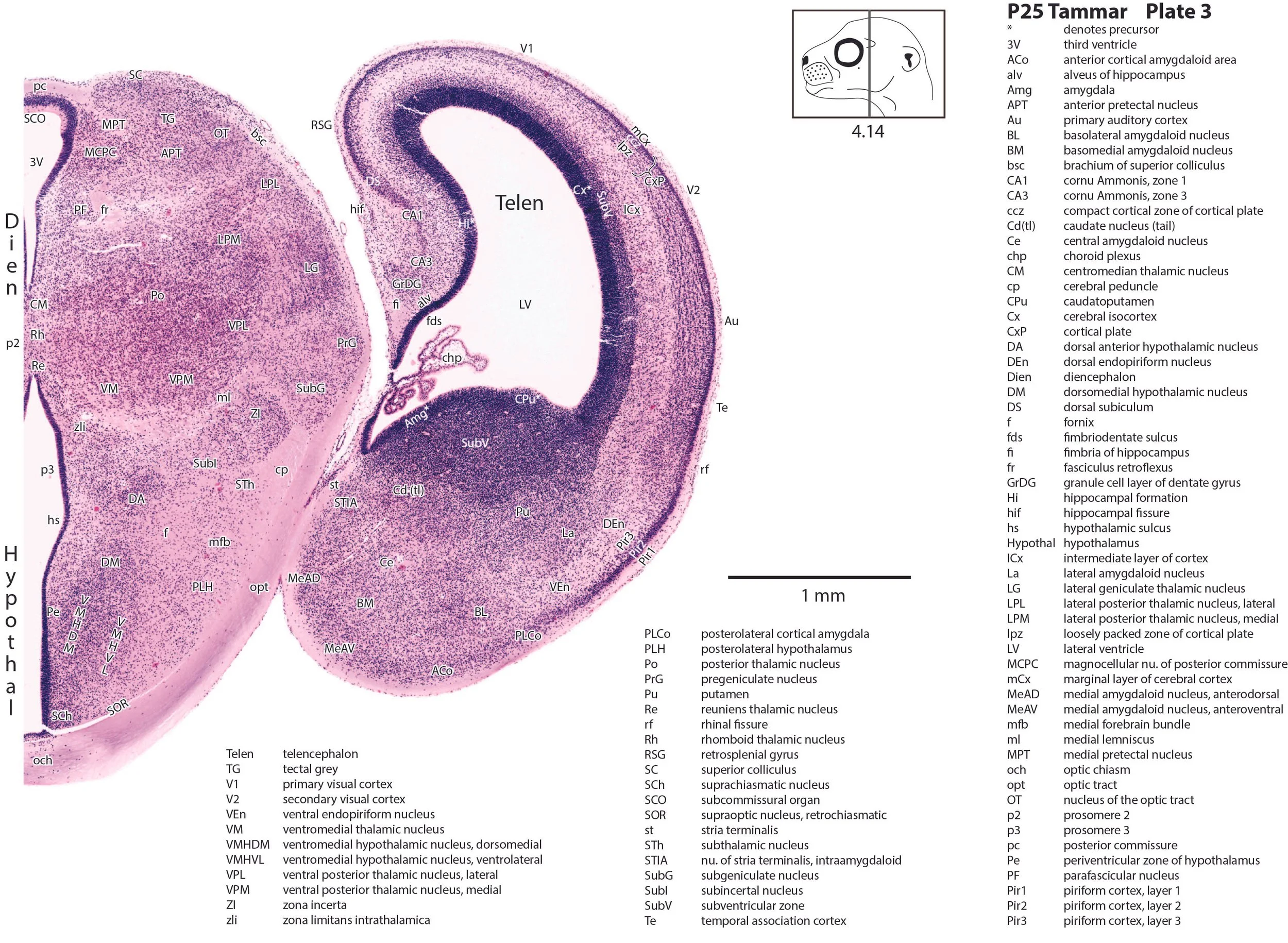
Each section was photographed with the aid of a Zeiss Axiophot photomicrographic system and a Canon EOS 400D body. Images were placed in Adobe Illustrator 2023 and delineated. Developmental regions (i.e. neuroepithelium) destined to give rise to adult structures have been denoted by the adult structure’s name with an asterisk (e.g. Cb* denotes the cerebellar developmental field of the rhombic lip).
The brain is essentially bilaterally symmetrical, so only half the brain has been depicted, with a 1 mm scale bar indicating the size of structures in the dehydrated tissue. A small finder cartoon has been provided in each plate to show the rostrocaudal position of the section. The distance from the rostral tip of the brain to the section depicted is given for each image.
Observations
The olfactory bulb now has discrete layers (glomerular layer – Gl; external plexiform layer – EPl; mitral cell layer – Mi; internal plexiform layer – IPl; granule cell layer – GrO; see Plate 1, 0.14 mm), but the olfactory bulb neuroepithelium (OB*) is still active, indicating the ongoing production of microneurons.
The cerebral cortex (see Plates 1 to 4) consists of an inner neuroepithelium (Cx*) surmounted in turn by a proliferative subventricular zone (SubV), an intermediate zone (ICx), a cortical plate (CxP) divided into a loosely-packed zone (lpz) and compact cellular zone (ccz), and finally a cell-sparse marginal zone (mCx). Distinct layers within the piriform cortex are visible (Pir 1, 2, 3; Plates 1 to 3).
The distinctive fasciculus aberrans (fa) of diprotodontid marsupials is now large (Plate 2). This bundle allows commissural axons from one hemisphere to pass from the internal capsule to the anterior commissure. Nevertheless, commissural axons from the lateral cortex (inferior frontal and temporal, including piriform cortex) continue to use the external capsular (ec) route to reach the anterior commissure (Shang et al., 1997).
Differentiation of subpallial regions within the telencephalon has begun, with the emergence of the neostriatum (caudate and putamen in Plate 2), bed nuclei of stria terminalis (STMV, STMD, STLV, STLD, STIA in Plates 2 and 3), ventral pallidum (Plates 1 and 2) and amygdala (Plate 3).
Discrete nuclei within the (dorsal) thalamus and prethalamus can be seen (e.g. LG, MG, VPL, VPM of the thalamus in Plates 2, 3)
The cerebellum has three deep cerebellar nuclei visible (Lat, Int, Med in Plate 7). Cerebellar cortical differentiation is slightly more advanced than at P19 with a loose Purkinje cell layer (Pk) and some granule cell precursors still positioned in an external granular (germinal) layer (egl in Plates 6 and 7).
Migration of neurons to the two major precerebellar nuclei (pontine and inferior olivary complex, Pn and IO) is still in process (see the migratory streams iom and pnm in Plate 8), but a substantial number of neurons have already settled in their final resting sites and distinctive subnuclei are emerging within the inferior olivary nuclear complex (see IOD and IOV in Plate 8).
References
Ashwell KWS, Marotte LR, Mai JK (2010) Atlas of the brain of the developing tammar wallaby (Macropus eugenii). In: The Neurobiology of Australian Marsupials. (K Ashwell, ed). Cambridge: Cambridge University Press.
Shang F, Ashwell KW, Marotte LR, Waite PM (1997) Development of commissural neurons in the wallaby (Macropus eugenii). J Comp Neurol 387: 507-523.