GAP-43 Immunoreactivity in the Head of the P0 Tammar Wallaby (Notamacropus eugenii)
Introduction
The tammar wallaby (Macropus eugenii) is a small macropod originally found in Western and South Australia and on ten or more offshore islands. Please see the introduction for the P5 H&E atlas for details of tammar wallaby biology.
GAP-43 is a calmodulin binding phosphoprotein, which serves as a substrate for protein kinase C and is thought to play a critical role in axonal guidance (Meiri et al, 1986). It is found in growth cones within both the peripheral and central nervous system and is a useful marker for developing axons. In these plates it has been used to show the position and trajectories of growing axons in both cranial nerves and central nervous system pathways (Hassiotis et al, 2002).
Methods
The wallaby depicted in the series of colour plates was obtained from a breeding colony at the Australian National University (ANU) in the Australian Capital Territory (ACT). Some monochrome images of this specimen have been shown in a previous publication (Hassiotis et al., 2002).
Please see the introduction to the P5 Tammar head GAP-43 sections for details about methods.
References
Hassiotis M, Ashwell KW, Marotte LR, Lensing-Hohn S, Mai JK (2002) GAP-43 immunoreactivity in the brain of the developing and adult wallaby (Macropus eugenii). Anat Embryol 206, 97-118.
Meiri KF, Pfenninger KH, Willard MB (1986) Growth-associated protein GAP-43, a polypeptide that is induced when neurons extend axons, is a component of growth cones and corresponds to pp46, a major polypeptide of a subcellular fraction enriched in growth cones. Proc Natl Acad Sci USA 83, 3537-3541.
Figure Legends
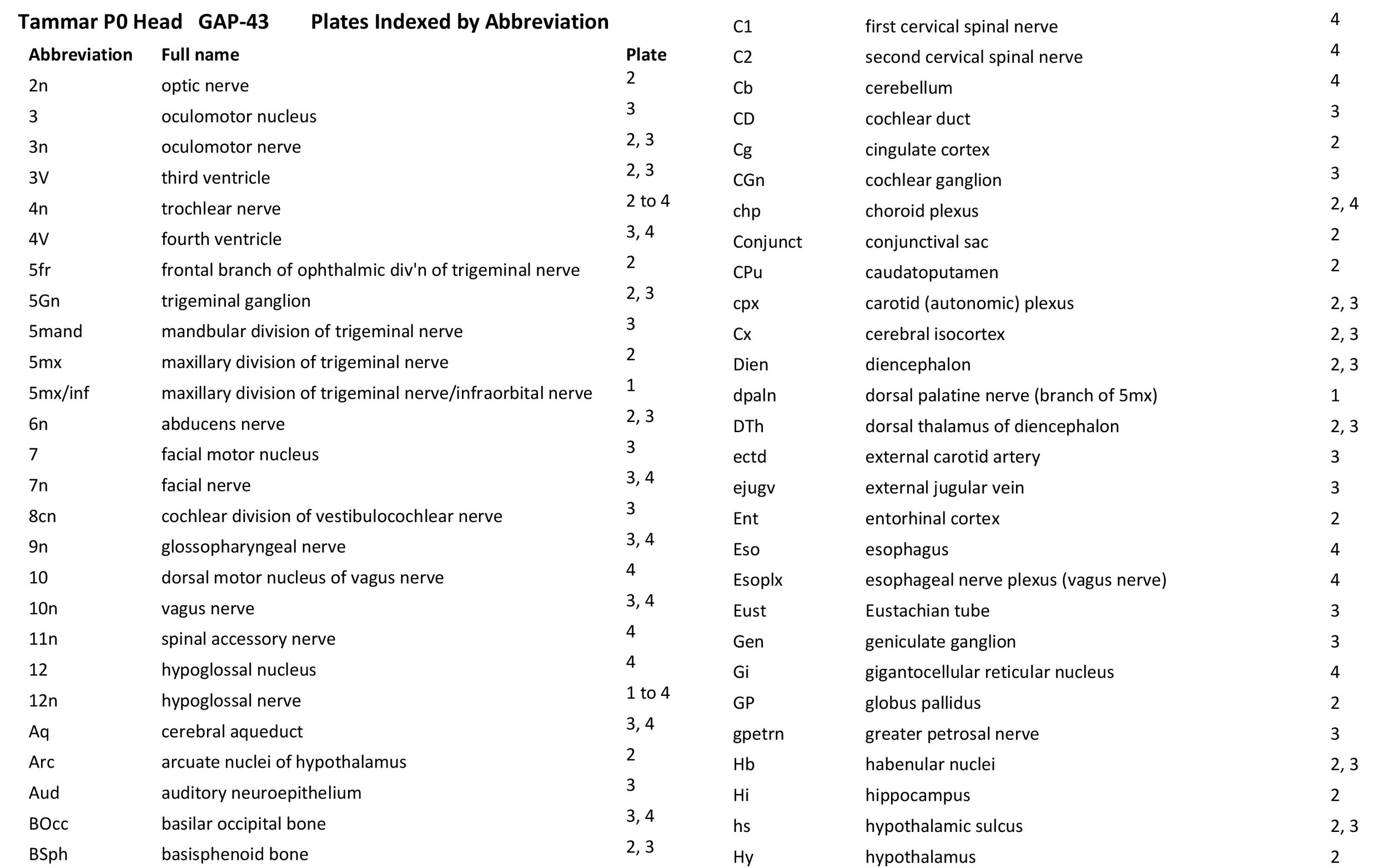
Figure 1 shows sections 212 and 232 through the head of a P0 tammar wallaby pouch young. These sections are respectively 2120 and 2320 µm from the tip of the snout, respectively.
Figure 2 shows sections 322 and 382 through the head of a P0 tammar wallaby pouch young. These sections are respectively 3220 and 3820 µm from the tip of the snout.
Figure 3 shows sections 412 and 472 through the head of a P0 tammar wallaby pouch young. These sections are respectively 4120 and 4720 µm from the tip of the snout.
Figure 4 shows sections 572 and 632 through the head of a P5 tammar wallaby pouch young. These sections are respectively 5720 and 6320 µm from the tip of the snout.


