Atlas of the Head and Thorax of an 8.5 mm GL Platypus (M37T)
Introduction
This series of images illustrates the extent of brain, spinal cord and vasculature development in an in ovo late pharyngeal arch platypus embryo.
A platypus embryo (stage KK) of 8.5 mm GL (greatest length) would be approximately 6 days before hatching (Ashwell, 2013), where hatching occurs at about 14 to 16 mm GL.
Details on platypus biology can be found at the Platypus page of this website.
Methods
The specimen illustrated here is M37T of the Hill collection, stored at the Museum für Naturkunde (MfN) in Berlin. The most likely site of collection is eastern New South Wales, near Sydney.
The specimen had been collected in October 1899 by JP Hill, embedded in paraffin wax and said to have been sectioned transversely (frontally) at 10 µm thickness, although the plane of section is almost horizontal. The stain was not identified on the specimen card, but it appears to be a variant of hematoxylin.
Images previously taken by KK Sulik and stored under open access within the MfN collection have been used for labelling (please see the MfN for access). Images were placed in Adobe Illustrator 2023 and delineated.
General observations
The vasculature of the specimen is in transition from embryonic to fetal architecture. The cardiac ventricles are incompletely septated and the truncus arteriosus is only incompletely divided in pulmonary trunk and aorta (TA in Plates 16, 17). A single aorta is present caudal to the heart, but paired dorsal aortae are seen rostral to the heart. Some pharyngeal arch arteries are still present (archa4 in Plate 13). The anterior cardinal veins (Lacardv, Racardv in Plates 16 to 19) are in transition to the mature venous pathways (SVC, brachiocephalic veins).
Telencephalon
The telencephalon consists of a thin pallium, made up mainly by a ventricular germinal zone and surrounded by only a thin preplate (PrePl in Plates 12 to 14), and small ganglionic eminences (see lateral ganglionic eminence – lge in Plate 12). Postmitotic cells are rare. The olfactory bulb (OB in Plates 14, 15) is primitive and unlaminated, with only an olfactory nerve layer present (ONL in Plate 15).
Hypothalamus
The hypothalamus consists almost entirely of the germinal compartments of the three segments (peduncular – hy1; terminal – hy2; and acroterminal – hyat; see Plates 8 to 12) with few or no postmitotic cells. Rathke’s pouch (Plate 10) and the infundibular recess of the third ventricle (InfR in Plates 9, 10) are present.
Diencephalon
The diencephalon consists of three prosomeres (from caudal to rostral - p1 or pretectum in Plates 8 to 11; p2 or dorsal thalamus in Plates 8 to 11; p3 or prethalamus in Plates 9 to 11). As seen for the telencephalon, postmitotic cells or neurons are rare. The epithalamus is thin and undifferentiated (EpiTh in Plates 8 to 12).
Mesencephalon (midbrain) and isthmus
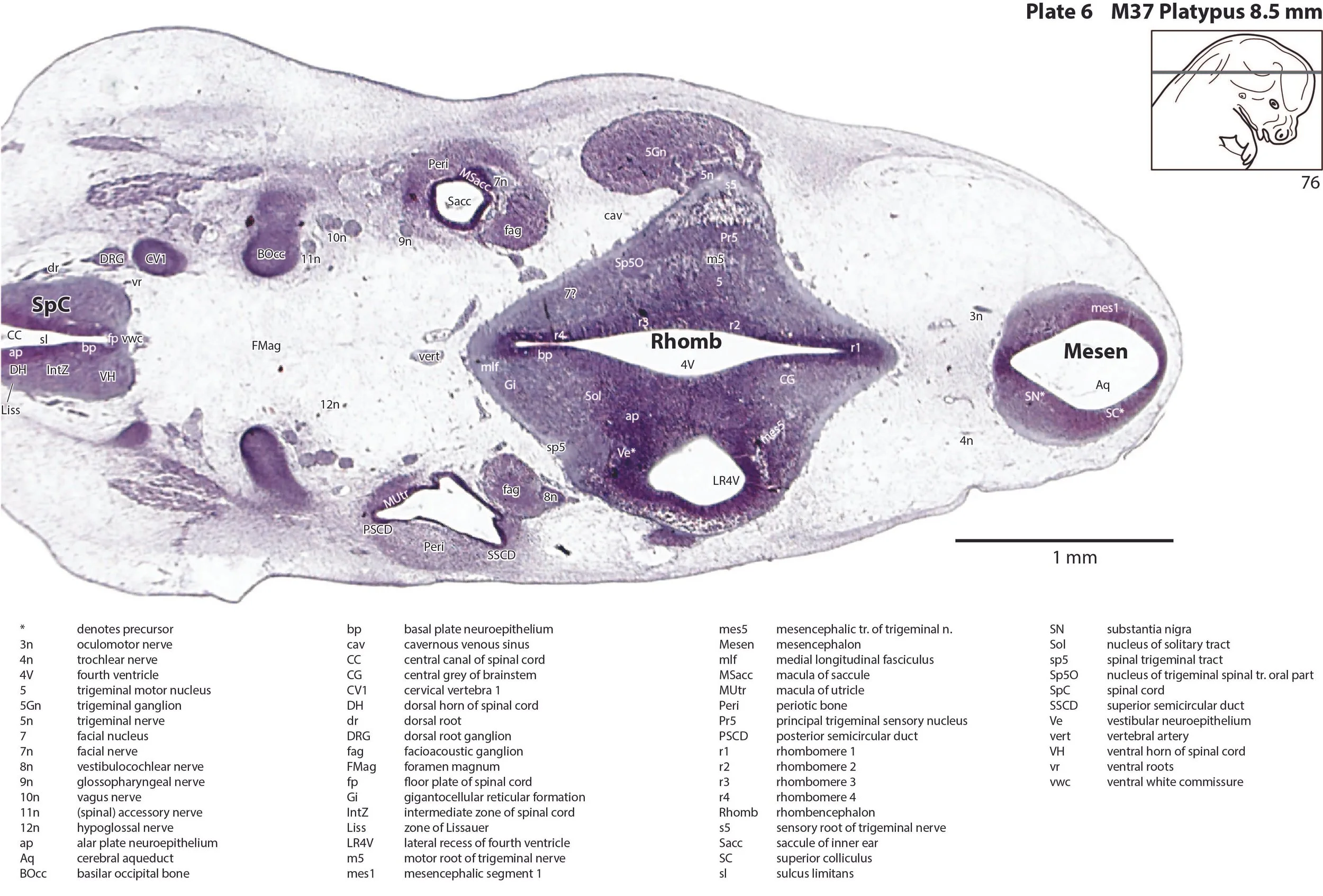
The midbrain is still immature and consists mostly of proliferative cell populations in the two mesencephalic segments (mes1, mes2) and isthmic segment (is). Some neurons have settled ventrally as putative substantia nigra (SN in Plates 6, 7), oculomotor nucleus (3 in Plate 5) and trochlear nucleus (4 in Plate 4).
Rhombencephalon terminology
The classical division of the rhombencephalon (hindbrain vesicle) into metencephalon and myelencephalon is not strictly valid given modern concepts of rhombomeric subdivision based on gene expression (Watson, 2012), so only a rhombencephalon (Rhomb in Plates 1 to 7) has been listed here. Where possible rhombomeres 1 to 8 have been indicated.
Rhombic lip
The rhombic lip is a developmentally important proliferative region at the margin of the fourth ventricle. The rostral rhombic lip (rrl in plates 3 to 5) is an important source of microneurons for the cerebellum, whereas the caudal rhombic lip (crl in plates 5 to 5) produces microneurons for the ventral brainstem (e.g. for Pr5 and precerebellar nuclei).
Cerebellum
The developing cerebellum (Cb in Plates 1 to 5) arises from the rostral rhombencephalon (r1 segment). It is very immature at this stage with only a few postmitotic cells (neurons) in presumptive cortical and nuclear transitory zones.
Ventral rhombencephalon derivatives
The ventral rhombencephalon has the largest population of neurons of any brain region, but differentiation of nuclear groups is limited. Alar and basal neuroepithelial components are visible (ap, bp in Plates). Some components of the trigeminal sensory complex can be seen laterally (Pr5 in Plates 6, 7; Sp5O in Plates 3 to 6; Sp5I in Plates 3 to 5) and a gigantocellular reticular formation (origin of descending reticulospinal connections) is present medially (GI in Plates 3 to 6).
Inner ear
The inner ear otic vesicle (Otic in Plates 3 to 5) has developed endolymphatic duct and sac extensions (ELD, ELS in Plates 2 to 4), semicircular ducts (PSCD, SSCD in Plates 4 to 6), utricle (Utr in Plate 5), saccule (Sacc in Plates 6, 7), and cochlear duct (CD in Plates 7, 8).
Cranial nerves
Cranial nerves 3 (oculomotor) and 4 (trochlear) can be traced from the mesencephalon and isthmus (respectively) forward (oculomotor in Plates 6 to 11; trochlear in Plates 2 to 11). Cranial nerve 6 (abducens) is seen in Plates 7 to 9. Cranial nerve 7 (facial) can be traced through the middle ear cavity into the second pharyngeal arch (Plates 4 to 10). Cranial nerve 8 is seen in Plates 3 to 6. Cranial nerves 9, 10 and 11 (glossopharyngeal, vagus and accessory, respectively) are found alongside the brainstem before emerging from the chondrocranium. The glossopharyngeal nerve (Plates 3 to 11) is best found adjacent to the sole muscle it supplies, the stylopharyngeus (Plate 11). The vagus nerve (10n in Plates 3 to 19) can be traced through its superior vagal ganglion (S10Gn in Plates 8 to 10) and is closely associated with the common carotid artery and internal jugular vein. The (spinal) accessory nerve (Plates 3 to 12) arises from the most caudal medulla and rostral spinal cord and passes forward to lie alongside the vagus nerve. It is easiest to identify outside the chondrocranium alongside the two muscles it supplies, sternomastoid (StM in Plate 11). The hypoglossal nerve (12n in Plates 4 to 12) can be traced into the intrinsic tongue musculature.
Spinal cord
The spinal cord grey matter is poorly differentiated and the white matter is very thin. The ventral horn is larger than the dorsal horn (VH and DH in Plates 4 to 19). A ventral white commissure (vwc in Plates 4 to 19) is present for decussating spinothalamic pathways.
Limbs and peripheral nerves
Nerves of the brachial plexus have penetrated beyond the base of the upper limb and some nerves (radn in Plates 15 and 16, medn in Plates 16 to 18) are visible near the humerus. The nerves of the lumbosacral plexus (not shown) haven’t penetrated beyond the base of the lower limb.
Acknowledgements
I would like to thank Dr Peter Giere of the MfN, Berlin Germany, for access to the collection and for all his kind help during the work.
References
Ashwell KW (2013) Embryology and post-hatching development of the monotremes. In KWS Ashwell (Ed.), Neurobiology of Monotremes: Brain Evolution in Our Distant Mammalian Cousins (1st ed., pp. 31-46). CSIRO.
Watson CRR (2012) Hindbrain. In The Mouse Nervous System (Eds C Watson, G Paxinos and L Puelles) pp. 398–423. Elsevier, San Diego.


