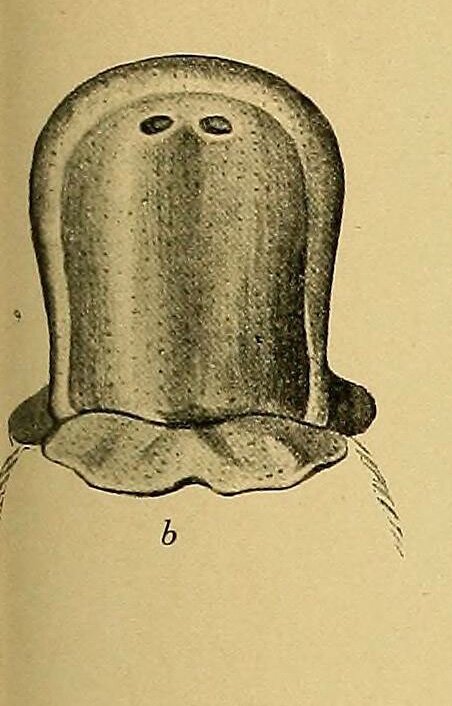
Platypus bill
See all Atlas of the bill of a newly-hatched platypus
The most remarkable sensory specialisation of the platypus is the concentration of electroreceptors and mechanoreceptors in the skin of the upper and lower bill (Scheich et al., 1986; Pettigrew, 1999) and served by the branches of the trigeminal nerve (see Figure 1a). These sensory gland receptors need moisture to function and the platypus operates in an aquatic environment, keeping the bill wet while feeding. Autonomic mechanisms allow the duct opening of the sensitive sensory glands (see Figure 1b) to close when the platypus is out of the water on the river-bank or in its burrow (Manger et al., 1998).
The platypus appears to use its bill as an antenna for electrical teloreception (i.e. sensation at a distance), whereas the echidnas use their beaks as push-probes for moist leaf-litter and soil. When swimming, platypuses move their bill to the left and right so that the electrical fields from the muscular activity of prey sweep across the sensory receptors. This allows the platypus to home in on the sources of electrical signals and use the mechanoreceptors to locate the prey by water displacement and direct touch.
References
Bethge P, Munks S, Otley H and Nicol S (2003) Diving behaviour, dive cycles and aerobic dive limit in the platypus Ornithorhynchus anatinus. Comparative Biochemistry and Physiology. Part A. Molecular and Integrative Physiology 136, 799-809.
Cabrera, A (1919) Genera mammalium Madrid, 1919. biodiversitylibrary.org/page/32474795
Grant TR (2007) The Platypus. 4th edn. CSIRO Publishing, Collingwood.
Manger PR, Keast JR, Pettigrew JD and Troutt L (1998) Distribution and putative function of autonomic nerve fibres in the bill skin of the platypus (Ornithorhynchus anatinus). Philosophical Transactions of the Royal Society. Series B. Biological Sciences 353, 1159–1170.
Pettigrew JD (1999) Electroreception in monotremes. Journal of Experimental Biology 202, 1447–1454.
Scheich H, Langner G, Tidemann C, Coles RB and Guppy A (1986) Electroreception and electrolocation in platypus. Nature 319, 401–402.

Platypus beak dorsal view modified from Cabrera, (1919) biodiversitylibrary.org/page/32474795

Ventral view Platypus beak modified from Cabrera, (1919) biodiversitylibrary.org/page/32474795
Fig. 1. a) Dorsal (superior) surface of the upper bill of the platypus showing large nerve bundles of the infraorbital nerve [branch of maxillary division of the trigeminal nerve; 5mx(inf)] innervating the electro- and mechanoreceptors. b) close-up of the dorsal bill surface near the nostril (naris) showing the pits of sensory gland openings.
![Fig. 1. a) Dorsal (superior) surface of the upper bill of the platypus showing large nerve bundles of the infraorbital nerve [branch of maxillary division of the trigeminal nerve; 5mx(inf)] innervating the electro- and mechanoreceptors. b) close-up of the dorsal bill surface near the nostril (naris) showing the pits of sensory gland openings.](https://images.squarespace-cdn.com/content/v1/60e638d550a959524454cb58/1628730205354-HBQ4RPP6U9NWHFA7DX8M/Fig1_Platypus_bill.jpg)